Showing posts with label physiology. Show all posts

Thursday, January 1, 2015

Your Metabolism Doesn't Know It's The New Year

This is a more scientific, detailed continuation of a discussion started on my other blog.  Fat Fun Fit: Why I'm Not Making Resolutions: A Case Against Dieting.  From that post (in blue):

How will your body react if your norm is to eat around 2,500 calories per day, and only a little walking as your physical activity, and you suddenly cut your food intake by HALF and jump up to a "perfect" workout regimen?  My body reacted like this, as would most:  "Holy crud!!  What the hell is going on?!!  We must be starving, and running to try to find food! What apocalypse is going on out there?  Oh man, this is it, this is the end.  Emergency!  Emergency!  Going to code red- crisis management mode!"  My body senses a sudden catastrophic world event on many January 1st's.

Now time for the science nerd in me to come out.  What happens physiologically while on a low-calorie diet that your body is not used to, is that all food coming in as much as possible will be stored as fat for later in case the emergency gets even worse, and muscle gets broken down to be used for quick energy.  Cause if you're in a crisis, keeping your brain well-fed, and planning for the worst is top priority.  Your brain is not able to use energy from fat, and if you are starving your brain by eating a severely low-calorie diet, muscle is the quickest way to get energy to your brain.  Your body doesn't really have a way to tell how much fat it already has stored so that it can "cap" it at a certain amount, which is why you can get very morbidly obese people.  Your body will just keep making more fat when it is in these crises even though you already have a lot, or when there is a plain old excess of energy coming in.  Losing that muscle lowers my energy and makes sustaining this plan much more difficult.

Continuing on....

Your brain needs glucose.  Your body does too.  It's the energy that gets broken down into usable energy for your body, known as ATP.  You can also use other simple sugars like fructose, but those actually just get converted to glucose before being used to make energy.

Glucose can be stored in a number of ways.  The easiest way is as glycogen.  That is a ginormous molecule of glucoses linked together in a network.  Your liver and muscles store glycogen so that your body and brain have enough energy between meals.  They can only store enough to last for a few hours (between meals).  When there is no glucose in your blood from a recent meal and there is a need for energy, the glycogen gets broken down to glucose.

In the muscle glycogen, the glucose is used right there in the muscles so you can still walk around, type at your computer, chase your kids, etc. between meals.  The liver is the bank account for your brain.  The glycogen in the liver is broken down to send glucose into the blood for the brain to use.  Brain always has priority.  If the brain doesn't get enough, the body must supply.

When glycogen is gone, the body will go to the next source.  Muscle.  There are a lot of Amino Acids - building blocks of proteins - in your muscles, cause that's what muscles are made of- loads and loads of proteins.  Those proteins can then be made back into glucose to send to the brain so it has energy.  This process is called gluconeogenesis  (gluco=glucose, neo= new, genesis= make;  make new glucose).

When you are on a very low-calorie diet or starvation, your body will break down some muscle to feed your brain.  In the absence of readily useable glucose in the diet, the same happens.  This is why people on Atkins/ low carb diets lose a lot of weight quickly at the beginning.  They are starving their brains of glucose, so the muscle gets broken down.  Well, muscle weighs A LOT.  It is much more dense and heavy than fat.  It also takes a lot more water to metabolize muscle, so the majority of those pounds you are dropping on those diets are muscle and water.  Great if all you care about is the number on the scale.  Terrible if you care about your body composition, shape/ size, and actual health.

The other way for glucose to be stored is for PARTS of it to be put into fat. Glucose has 6 Carbons in it, but a bunch of 2-carbon subunits (Acetyl-CoA) can be put together into a long chain: a fatty acid.  Then 3 of these chains can be attached to a glycerol and stuck in adipocytes- fat cells.  A lot of energy can be tucked away this way.  You get loads and loads of energy out of fat.  You are probably familiar with this if you pay attention to nutrition labels.  There are 9 Calories per gram of fat, but only 4 Calories per gram of carbohydrate or protein.  That's equalizing the weight.  Fat takes up a lot more space, so all that extra energy stored in your body also makes your body bigger than the same amount of energy stored in muscles or glycogen.

So, how and when does this fat energy get used?  That's the tricky thing.  Your brain lacks the proper gateways and enzymes to metabolize the subunits from fatty acids (acetyl CoA) directly.  It can only take in glucose or ketone bodies.  The glycerol from the triglyceride can be made into glucose, so those get sent off to the liver to do that.  But the fatty acids are better used by the cells of the body cause they have the right enzymes and gates to allow that to happen.

But since fat is long-term storage, just like a trust fund, the body is not going to break into it at the first sign of trouble.  It waits to see if it can get by with the cash on hand, the checking and savings account.  (Glucose in blood from your meal, then glycogen, then muscle as explained already.)  Then if it's dire enough it will go for those fatty acids IF the body is in need of it.  So, from what I have heard from a Physiology professor about some research (but haven't located the research myself yet so I have no link, sorry), is that the mark for the fat getting mobilized is about 45 minutes of exercise.  After that point you would start to break down the fat for your body to use.  So the recommendation of 30 minutes of exercise most days doesn't even touch that.  If I want to reduce body fat, the best thing is to go on hours-long hikes as much as possible.  (The regular aerobic exercise IS crucial to your health in other ways though, and should still be maintained for your cardiovascular, respiratory, muscle and mental health.  Benefits of regular exercise are nearly endless.)

I mentioned the brain can use ketone bodies.  In severe starvation, the fatty acids can be made into ketone bodies, which can get into the brain and used for energy.  But they are very dangerous because they turn the blood acidic and it is detrimental to your body.  This is the last ditch effort during severe starvation, to keep your brain alive, cause without your brain, the show is over.

Thursday, September 4, 2014

Blood Brain Barrier

No one would argue blood is very important to our bodies!  It carries very important things to all parts of the body we need such as glucose and oxygen.  It also takes out the trash by removing wastes like lactic acid and carbon dioxide.

Blood is the highway by which our immune system cells gets around our body to take care of anything that invades.  Blood is also how medications, drugs, poison and toxins, hormones, etc. can get around our bodies.

But let's talk about blood and the brain.  Our brain is a very special organ that deserves special protection.  It's the only part of our body that is protected by a 7 mm thick covering of bone, in addition to cerebrospinal fluid cushioning and protective layers of meninges.  That protects from the outside in, but we also have protection from the inside out, called the blood-brain barrier (I will abbreviate it BBB).

Blood supply is very important to the brain so it has a constant supply of energy and waste removal.  Here are some diagrams showing the blood vessels supplying the brain.


Notice the arch at the bottom of this diagram is the aorta which comes right off the heart itself


The Common carotid artery is the one you are feeling when you take your pulse on your neck

This "Circle of Willis" shows the blood supply on the inferior/ ventral side of the brain.  You can see in the image on the right where this is in relation to the brain.


Alright, so we need that blood and it definitely is there.  But how to protect it?  Some may think the "Blood-brain Barrier" is some kind of a gate the blood goes through when it enters the vicinity of the brain, but that isn't the case.  There isn't a particular spot for the BBB, but rather, it exists as protection on the capillaries (smallest blood vessels where material exchanges happen) themselves in EVERY location within the area of the brain.  It's not a matter of filtering all the blood as it travels through your head, but it's a matter of being more selective about what things cross over FROM that blood into the brain tissue.

We have special gate-keepers to protect things from getting into our brains.  Here's a cross-section of what a blood vessel in the brain looks like compared to a regular one elsewhere in the body:


Not only are the capillary cells (red in the diagram) closed more tightly so things can't leak through, but the entire blood vessel is covered with the "feet" of astrocytes.  (My favorite glia!  Here's a post about them.)

Here's a more 3D view:


See how is it a gatekeeper?  Anything in the blood must go through the astrocyte in order to get to the neuron.  Astrocytes are like the bouncer, protective big brother, or best friend: "if you want to get to the neuron, you have to [quite literally] go through me first!"

Astrocytes are really integral to the chemical integrity in the brain and are a bit of the "unsung heroes" of the brain.  Not only are they gatekeepers, but they act as a kind of mop-up crew and storage unit for any leftovers the neurons leave around (like ions, some neurotransmitters), and they serve to make sure the neuron stays well-fueled, like a mother who keeps snacks in her purse for her toddler.  No wonder astrocytes far outnumber neurons in the brain.

How do these tight blood vessels and "feet" of the astrocytes actually protect it? They are cells, which means they are surrounded by membrane- a phospholipid bilayer, which looks like this up close:
Because of this configuration, stuff that is polar (charged) or water-soluble can't get through the membrane- it can't get past all those hydrophobic fatty tails. 

YOU SHALL NOT PASS!!!
Water-soluble stuff such as nutrients (Amino Acids, Glucose, vitamins)
Polar stuff
Chemicals & toxins
Viruses
Bacteria


Stopping viruses and bacteria for the win.  Stopping nutrients? FAIL.

So to fix that, we have special transporters to let the good stuff in.  They can be super specific, so a glucose transporter will ONLY let glucose in.

Okay, you can go in...
Non-polar/ uncharged/ fat soluble stuff: this includes oxygen going in and carbon dioxide going out
Drugs that are fat-soluble
Other important stuff with special transporters embedded into the membrane to let them in, like water, glucose, amino acids, vitamins, etc. (Glucose has a wicked-awesome backstage pass, AND it knows the lead singer of the band.)


Whew!  That's a big job and an important one for the BBB.

Stay curious!

Brain Development


It astounds me how much brain development takes place in a fetus before a woman even usually knows she is pregnant.  This is an important reason why many foods are fortified with folic acid.  Folic acid (or folate) is essential in early brain development to the point that in its absence, there can be severe defects (such as spina bifida), but by the time the woman discovers she is pregnant, damage is already done, because it affects the neural tube which has already developed by day 21 of pregnancy!

Quote from the Mayo Clinic:
"Spina bifida is part of a group of birth defects called neural tube defects. The neural tube is the embryonic structure that eventually develops into the baby's brain and spinal cord and the tissues that enclose them.
"Normally, the neural tube forms early in the pregnancy and closes by the 28th day after conception. In babies with spina bifida, a portion of the neural tube fails to develop or close properly, causing defects in the spinal cord and in the bones of the backbone."


The Spina Bifida example serves to show how quickly brain development takes off.  This video has an excellent animation of brain development in a human fetus.


A couple other visualizations of the neural plate becoming the neural groove and then neural tube:


Lastly, here's a nice TED talk to get you thinking about infants in a different light. thanks for sharing Claudia Lieberwirth.


Tuesday, August 26, 2014

Neuron Communication

I have blogged quite a bit about Action Potentials which are the way in which a nervous impulse travels down an individual cell. (For your reference: Action Potentials and Action Potentials Up Close).


Here I will summarize what it takes for a message to be passed from one neuron to another.  If you are interested in information about the specific channels that make neuron communication possible, see this post on Neurons.


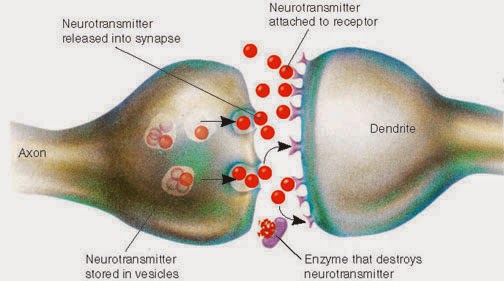
When an action potential - the electrical signals that neurons use to send messages - reaches the end of an axon, the electrical message must become a chemical message in order to cross the synapse. Neurons are connected to each other by a synapse which consists of the axon of the presynaptic neuron (the one sending the message), and the dendrite or cell body of the postsynaptic neuron (the one receiving the message), and the space between them called the synaptic cleft. Chemicals have to be sent across this space for the message to continue on. The chemicals used are neurotransmitters.

Here's a simple diagram of a synapse:


For a little more information and overview of neuron communication, here is a more detailed diagram:

When the electrical signal reaches the end of the axon, a special thing happens with Calcium entering the cell which allows those vesicles full of Neurotransmitter chemicals to exocytose.  That's a fancy word meaning the vesicle smooshes into the cell membrane, kicking its contents outside the cell- into the synapse.  Those neurotransmitters are then free to act on the postsynaptic cell dendrites or body to continue the message along!

But how did those vesicles of neurotransmitter actually GET there?  The cell factories that make neurotransmitters which are mostly made of protein, are waaaaaay back at the cell body, but it has to be released at the terminal button of the axon!  Axons can be very long, like so:

Neurotransmitter vesicles are continually made by the neuron (each neuron only makes one particular type of neurotransmitter), and sent down the axon.  They get there the same way you would travel a long distance- on a "highway" of sorts.

Here is a great animation of the whole neuron communication process, and you will see vesicles coming down to the end of the axon.


So, the cool thing is those vesicles are literally "walked" down the microtubule highway.  In my favorite video, you can see this happen:


Lastly, I found this funny little video about the life of a motor protein (the alien-looking guy that walked the vesicle down the microtubule).  Enjoy!


P.S.  While searching for images, I ran across this amazing blog, so here is a great reference on the brain and neuroscience, explained in simple terms in much the same way I try to do my own blog. The Brain Geek

Stay curious!
-Julie

Friday, April 4, 2014

Blood Clotting

Blood clotting or coagulation (also called hemostasis), is complex, but we want to try to understand this in "big picture" form first off.

When an injury occurs in a blood vessel, here are the steps we go through.


Classes.midlandstech.com

Platelets and Fibrin are the important things that we actually get as products of coagulation that seal up a wound.  But the activation of them is a complicated process.  Which is a good thing for your body!  If it were an easy reaction, we could get our blood spontaneously clotting on us and that would be BAD.

Here's another way to visualize this:

studyblue.com

In first forming the platelet plug, it's important to note that it's the exposed collagen fibers that are attracting platelets.  The normal, healthy state of things is to NOT have platelets sticking to blood vessels.  The ability to not stick is ensured by the blood vessel lining cells (endothelial) releasing prostacyclin to prevent platelet sticking.  But with damage, that isn't released and instead collagen causes platelets to stick.  This can be visualized here:

dc416.4shared.com

Now for the actual clotting part after the platelet plug, for now I have some videos.  If you want to cut right to the chase and a great explanation, watch the last video.


Videos
Here's a not fabulous animation but an animation nontheless...


This short clip I just found helpful to visualizing how the different factors work together to activate factor X then prothrombin, to activate thrombin which they refer to as the "thrombin burst" because it creates a large amount then creates a positive feedback loop to further increase the effect.


This is the best video I found!  I like how concise and understandable he makes it, looking at the big picture and working backward from there.  Hope it's helpful for you too.

All clotting factors are made by the liver, except 8 and PAF3. (Platelet activating factor is made by white blood cells.)

Saturday, November 16, 2013

Neuron Ion Channels - Detailed

Inward rectifying potassium channels.  This is what started all this.  Just a little paranthetical aside in my Neuroscience teacher's notes that left me curious.  One thing led to another, and here we are.  Ion channels.  Hope you learn something like I did.  Enjoy!

A plethora of potassium channels
Here is a short video showing the molecular structure of a Potassium channel and how it can be perfectly selective to allow Potassium and not Sodium through.

And look at this beautiful top view of a potassium channel with a little purple potassium ion in the center.


Inward-Rectifying Potassium Channels
What does inward-rectifying mean?  Simply put, it just means that voltage travels inward more easily than outward.  So this name tells us that Potassium will move into a cell when open.
(Example - cardiac muscle cells- responsible for the long refractory period between beats, to avoid tetany; kidneys, regulating potassium ions

Action Potentials: Delayed Rectifier Potassium Channels & A Type Channels (outwardly rectifying)


Tandem Pore Domain Potassium Channels (Leak Channels)

Voltage Gated Potassium Channels


********This post under construction!  I just found a textbook with an entire chapter on ion channels, so after I study for my test I will dive into this and finish the post...  thank you for not hating me too much for leaving you hanging.  You'll just have to subscribe to learn more! :)

Tuesday, October 1, 2013

Bio Chemistry in Plants

I love when multiple classes overlap in what they're teaching me.  Today it's Bio Organic Chemistry and Plant Biology.

Top half of the diagram below:
This is showing glucose.  At first it's in open chain form.  Then it becomes cyclized by forming a hemiacetal - a bond between the carbonyl on Carbon 1, and the alcohol on Carbon 5, which is where we get the Oxygen on the top right corner of all cyclized glucose molecules (was the carbonyl attached to Carbon 1).


Bottom half of the diagram:
This shows how the new hemiacetal can be twisted one way or another so that when the bond is made, the alcohol from carbon 5 is either facing up or down off of its new home on Carbon 1.  The glucose on the left shows the OH on the bottom, so it's considered the alpha form.  The glucose on the right shows the OH on top, which is the beta form.  (Memory Aid: Alpha= the letter looks like a fish, which swim below the surface so the OH is pointed downward, Beta= "B" is for bird, which flies above the surface so the OH is above.)


So we have 2 forms of D-Glucose in biology.  Now we can see an example of how they can be put together into polymers in plants.

Starch
Starch is a string of glucose attached by alpha glycoside bonds.  I made some little drawings to walk us through how these look.

1) We start with two alpha-D-glucose molecules next to each other which looks like this:

Notice they are both in the alpha configuration because the OH is on the bottom for Carbon 1. Ooops forgot to label the Carbon numbers until later, sorry.  Carbon 1 is the one on the right end of each hexagon.)

2) Now we will bond these together using dehydration synthesis by removing an OH hydroxide from one side, and just the H from the hydroxide group on the other side.  This becomes a water molecule which is a byproduct.  We took water out so that's why it's called dehydration synthesis.

3) So now we can see that these two glucose molecules are bound together by the one Oxygen that was left over.  This is an alpha-1-4-glycoside bond.  The 1 and 4 are because it's between Carbon 1 on the glucose on the left, and Carbon 4 of the glucose on the right, as I circled on the picture below, with the now labeled Carbon numbers. :)


 4) Make this bond many times, and you will get a big starch chain held together by many alpha glucose bonds, like so:


Cellulose
Cellulose is put together differently because it uses Beta-D-glucose.

1) Two Beta-D-Glucose molecules next to each other, and we could pull out the water molecule from this like so:

2) But notice that it's a little awkward, so instead we flip one of the glucose molecules upside down, which you can tell by the H2COH group being on the bottom of the glucose on the right.


Now the OH's match up better so we can make the glycoside bond.

3) Here's our brand new Beta Glycoside bond, more specifically a Beta-1-4-Glycoside bond


4) And once again we can make a nice chain of these, and you can notice how every other glucose molecule is flipped upside down.


There's our cellulose!  Enjoy, hope you learned something, and I hope my drawings were clear enough to make sense.  Stay curious!


P.S. Fun side note - this is my very first Bio/ Organic Chemistry post on the blog!  Have to add a new label to the cloud..woohoo! :)

Wednesday, April 17, 2013

Kidney Physiology


Great overview


Urine Production Video - Osmotic Gradients

This is kind of fast, but it shows the counter-current diffusion of water and ions from the Nephron loop and vasa recta.

.


Here's a fun video - why coffee and alcohol make you pee more.


Wednesday, April 10, 2013

Respiration, Oxygen, and Hemoglobin

By decreasing pressure inside the thoracic cavity, we are able to inspire air.  By contracting the diaphragm, intercostal muscles, and others, we increase the size of the thoracic cavity, which in turn lowers the pressure to the point that it is lower than the outside air, causing a pressure gradient so the air rushes into the lungs.  To exhale, it is the opposite- relax the muscles, decrease the space in the cavity, which increases the pressure inside until higher than outside, so the air rushes out to where the pressure is lower.

This video is a good summary.  The embedding doesn't work, so you'll have to click the link instead.  Just left the embed on there for the picture. :)

Partial Pressure
Total Pressure of air can be broken into the partial pressures of all the gases contained in that air.  For instance, if the atmospheric pressure is at 760 mm Hg, and 20% of that is Oxygen gas, 80% Nitrogen gas, then the partial pressure of O2 would be 20% of 760 = 152 mm Hg.  Partial Pressure of N2would be 80% of 760 = 608 mm Hg.
This illustrates how increasing the pressure, as in the B picture, causes more gas to diffuse into the liquid.  A simple way to think about it is just that the higher the partial pressure of that gas, the less space it has to bounce around in the air, so more of it will end up in the liquid.

Here's a long video on partial pressure and gases getting into solution, I didn't watch the entire thing yet but it looks like a good detailed explanation for those who feel they could use more information.

Friday, March 22, 2013

Vision - colors

This podcast about color is so awesome.


Wish I could have the eyes of a mantis shrimp!  We have only 3 types of cones (photoreceptors) for color vision, they have SIXTEEN!  I always feel like there must be colors that I can't see and wonder what they would look like.  I will be coloring a picture and looking around for different colors to use and I just can't find them and I realize that's all that I can see.

Tuesday, March 5, 2013

Muscle Contraction

Muscle Contraction is a very cool, amazing process.

Within the myofibrils, this is what's happening:
Source
Here's the explanation of the picture from the source's website:
a. Without calcium ions (in relaxed state). Because tropomyosin completely covers the sites (indicated in blue) of actin binding to myosin, myosin cannot bind to actin.
b. When calcium ions released from excited muscle cells bind to troponin, the binding sites of actin are exposed owing to the shift of tropomyosin; however, the extent of the shift is not sufficient for myosin to bind to actin.
c. When small amounts of myosin heads bind to the binding sites of actin under state b, tropomyosin shifts further, resulting in the complete exposure of the binding sites of actin.
d. Because the binding sites of actin are completely exposed, many myosin heads can bind to actin, which enables the generation of a large muscle contraction power. 


I love that it is showing the myosin heads (green globs) all the way around the actin.  Most of the time we look at it as a very 2-dimensional thing, but when you look at the picture below, you can see that it is a very 3-dimensional process, with myosin heads able to bind all the way around.  Each of the small dots represent actin, the bigger ones are myosin (seen on the ends of the myofibrils).
Source

Keep in mind, myofibrils are organelles within a muscle cell.  So the sarcoplasmic reticulum is the muscle version of ER, wrapped around each myofibril.  Stuff is diagrammed 2D to make it simpler and more understandable.  Once you understand it, it's cool to look back and imagine the bigger picture.


Videos!

Love to weed through the options and find a few good ones for you, this time I've included a song, rap, and skit!


Very short and sweet, this is just a good little animation to show how the filaments slide past each other, with the myosin heads moving along at different times.

One recommended by a student.  It says it's for invertebrates, but looks like it's accurate for us as well.


Hahaha, this is a fun one.  A song/rap explaining the whole process.  I probably like it because the background music is Daft Punk (they did the music for Tron Legacy).  Anyway!  Maybe it's helpful for someone, and it's kinda fun to listen to.


This skit is a great idea.  It's hard to read their signs some times, but this was a fun little video and it might help a couple things click for you like it did for me.  I want to get a bunch of people to act something like this out some day! :P  (Don't worry, no plans to make my study sessions do this...)


The singing on these videos often leaves much to be desired!  But the info in this one is pretty good...

Here's Dr. Ashworth's explanation she gave permission to share.  Thanks Jessie for recording and sending it.


Enjoy. :)

Wednesday, February 27, 2013

Nervous System

The nervous system is divided into Central Nervous System (brain and spinal cord) and Peripheral Nervous System (nerves, ganglia, etc).
Here's a little chart, courtesy of a great Physiology student:

Peripheral Nervous System Central Nervous System
nerves tracts
ganglia nuclei
Schwann cells oligodendrocytes


Part of the Peripheral Nervous System (PNS) is the involuntary part, consisting of efferent neurons controling visceral organs.  This is called the visceral nervous system, but more commonly known as:
Autonomic Nervous System
Autonomic = Automatic = Involuntary
Sympathetic & Parasympathetic divisions.  Here's a silly little video that shows the functions of each.


Sympathetic Division
Location: Thoracic and Lumbar regions of spinal cord

3 Kinds of pathways


Parasympathetic Division
Location: Brain, and Sacral region of spinal cord


Receptors


This is super fun, talks about the brain, gets deep and slightly philosophical, and mentions reflexes.  It's all about the brain! :)


Monday, February 18, 2013

Reflexes

Reflex Arcs involve the spinal cord and leave the brain out of the picture, in order for the compensatory action to happen very quickly and avoid tissue damage.  Here is a short and sweet explanation:


Thursday, February 14, 2013

Glucose Metabolism - Glycolysis through Kreb's Cycle

I was trying to think of a way to model these processes for the benefit of kinesthetic learners, so this is what I came up with using these cool toys my kids got from the kids' meal at Wendy's.  I hope this will help people visualize this and understand where all the Carbons from glucose go, and why on earth we care about all those NAD, NADH, FAD, and FADH2.


Here are the symbols for this model.  Purple= Carbon, Blue= Hydrogen, Red=NAD, Green=FAD, Orange=CoA enzyme


Below is a VERY simplified version of a glucose molecule.  This shows the 6 carbons because we want to see what happens to all these carbons.  There should be 12 Hydrogens and 6 Oxygens as well but for simplicity, I have only the carbons and 2 Hydrogens to illustrate one round of Redox reactions in glycolysis.

Glycolysis
Here we start with a glucose molecule and we will break it down.  For this demonstration I am ignoring the ATP, but just note that 2 ATP's go in to Glycolysis and you get 4 ATP out (so, a net production of 2 ATP).

Here are the reactants, or what goes in to glycolysis:


Next, we see that the glucose is broken apart (in half, basically)

And the NAD's come in to take the Hydrogens off to become NADH.  FYI, the glucose was "oxidized" because the electrons were taken off it, and the NAD was "reduced" because it got the electron (the hydrogen).  So, this is a Redox (Oxidation-Reduction) reaction.

Now we have two 3-Carbon structures, and two NADH's


The 3-Carbon moleucles are Pyruvate.

That's the end of glycolysis.  The NADH's are sent on to the electron transport chain at this point, so they leave.  Next the Pyruvates will be taken on and processed further in the next process, sometimes called the "Linking Step".  We will follow one pyruvate - keep in mind this process would happen twice for each glucose molecule since it splits in half into 2 pyruvates.

Oxidation of Pyruvate to Acetyl CoA (aka "The Linking Step")
So here is the Pyruvate going into the linking step.  By the way, this is the point at which metabolism has entered the Mitochondria.  Glycolysis takes place in the cytoplasm, and then pyruvate enters the mitochondria and that is where the rest of the processes take place.


I didn't do an intermediate picture on this one, sorry about that.  The NAD comes and harvests another Hydrogen (which is not shown on my simplified pyruvate), CoA is added and one carbon is removed and disposed of as Carbon dioxide.  Here are the products, notice the 3 Carbons accounted for:


And, we see here where each of the products are headed now:

Kreb's Cycle
Then we follow the Acetyl CoA into the Kreb's Cycle, here are the reactants:


I didn't attempt to show all the steps of the Kreb's cycle here, but there are many redox reactions taking place, turning NAD and FAD into NADH and FADH2.  Here's a diagram of the cycle, if you really want to see it:


 Here you see the products, and see all the carbons are accounted for (2 Carbons in, 2 Carbons out):


And, here are the other products, which are the electron carriers that then deliver it over to the electron transport chain:

Also, note that 1 ATP is made during the Kreb's cycle which is not illustrated.

See this post on the Electron Transport Chain for some great videos about what happens there.  NADH and FADH2 are the carriers that drop off the electrons to run the electron transport chain, which harvests 30+ ATP per glucose molecule.  That is why we care so much about harvesting Hydrogens from glucose so we can make NADH and FADH2 and send them on their merry way to the electron transport chain to make ATP.  ATP is the energy that cells use to do pretty much everything!  If you stop making ATP, you die.  End of story.


So, to sum up - see if you can go through and account for all the Carbons, Hydrogens and Oxygens of glucose (glucose is C6 H12 O6).  You'll see that 6 carbons go in, and 6 carbons come out.  (Remember that each glucose molecule becomes TWO pyruvates, so you would double the numbers from that point on.)

Also, 6 oxygens go in, and how many come out?  Yup, 6.  Go back and check if you want to see for yourself. :)

Now, how about the Hydrogens?  Go through and count to see how many are harvested.  Really, go look and figure it out...


....
Did you come up with 14?  Did you count wrong?  NO!  It is 14.  But there are only 12 on glucose right?  What's the deal?  The reason is we actually have one molecule of water (H2O) that goes in the Kreb's cycle during one of the steps (it's added to fumaric acid to make malic acid), and those are then harvested off.  So there are the extra 2 hydrogens!  :)  It's a beautiful thing.

P.S. if you notice on the diagram of Kreb's, it shows 2 H20 going in, but one of them does come back out, as shown on the more detailed diagram below.  2 water molecules in, 1 back out, that's a net of 1 H20, so there are your extra 2 Hydrogens still.  I circled the H20 for you to see:
Source

Hope that helps you understand and visualize metabolism better!  I'd love if you would leave a comment, and follow this blog if you feel so inclined.  Thanks, and happy learning. :)