(This post has resources and information for learning about Memory and Amnesia, for Physiological Psychology.)
Radio Lab:
6:30 Rat tests, 8:08 Chemical prevent memory in rats; Clive Waring 42:30
The Hippocampus and Patient H.M.(by Ted Ed)
H.M. - Nova special
9:22 What like for H.M. 10:40-11:52 Muscle memory star test
Morris Water Maze
Showing posts with label physiological psychology. Show all posts
Showing posts with label physiological psychology. Show all posts
Thursday, October 30, 2014
Thursday, September 4, 2014
Directional Terms - human
Here is a handy reference on directional terms used for human anatomy. I was nice enough to choose the pictures of people with clothes on. You're welcome.


Here are some specific to the brain, because for humans our brain as we are standing is tilted compared to the way we hold up our heads, like so:
So simply using superior/inferior and anterior/posterior doesn't quite work for how we normally think of orienting the brain, which is why we use dorsal/ventral and rostral/caudal.

Here is a memory aid for this. I had a hard time keeping dorsal/ventral straight, so this is what helped me.
When I think of dorsal, a shark comes to mind with its iconic dorsal fin on its back or in this case top:
.jpg)
For ventral being the bottom or down, I had to think about stingrays. They take water in on the top of their bodies and then shoot the water out the bottom over their gills. So they vent the water out the bottom side of their body. Hope that helps.
There ya go, have fun in anatomy or whatever class brought you to find this blog!
Stay curious.
Here are some specific to the brain, because for humans our brain as we are standing is tilted compared to the way we hold up our heads, like so:
 |
| Source on Studyblue |
So simply using superior/inferior and anterior/posterior doesn't quite work for how we normally think of orienting the brain, which is why we use dorsal/ventral and rostral/caudal.
Here is a memory aid for this. I had a hard time keeping dorsal/ventral straight, so this is what helped me.
When I think of dorsal, a shark comes to mind with its iconic dorsal fin on its back or in this case top:
For ventral being the bottom or down, I had to think about stingrays. They take water in on the top of their bodies and then shoot the water out the bottom over their gills. So they vent the water out the bottom side of their body. Hope that helps.
 |
| Left image: top/ dorsal side of stingray. Right image: bottom / ventral side of stingray |
There ya go, have fun in anatomy or whatever class brought you to find this blog!
Stay curious.
Blood Brain Barrier
No one would argue blood is very important to our bodies! It carries very important things to all parts of the body we need such as glucose and oxygen. It also takes out the trash by removing wastes like lactic acid and carbon dioxide.
Blood is the highway by which our immune system cells gets around our body to take care of anything that invades. Blood is also how medications, drugs, poison and toxins, hormones, etc. can get around our bodies.
But let's talk about blood and the brain. Our brain is a very special organ that deserves special protection. It's the only part of our body that is protected by a 7 mm thick covering of bone, in addition to cerebrospinal fluid cushioning and protective layers of meninges. That protects from the outside in, but we also have protection from the inside out, called the blood-brain barrier (I will abbreviate it BBB).
Blood supply is very important to the brain so it has a constant supply of energy and waste removal. Here are some diagrams showing the blood vessels supplying the brain.
This "Circle of Willis" shows the blood supply on the inferior/ ventral side of the brain. You can see in the image on the right where this is in relation to the brain.
Alright, so we need that blood and it definitely is there. But how to protect it? Some may think the "Blood-brain Barrier" is some kind of a gate the blood goes through when it enters the vicinity of the brain, but that isn't the case. There isn't a particular spot for the BBB, but rather, it exists as protection on the capillaries (smallest blood vessels where material exchanges happen) themselves in EVERY location within the area of the brain. It's not a matter of filtering all the blood as it travels through your head, but it's a matter of being more selective about what things cross over FROM that blood into the brain tissue.
We have special gate-keepers to protect things from getting into our brains. Here's a cross-section of what a blood vessel in the brain looks like compared to a regular one elsewhere in the body:

Not only are the capillary cells (red in the diagram) closed more tightly so things can't leak through, but the entire blood vessel is covered with the "feet" of astrocytes. (My favorite glia! Here's a post about them.)
Here's a more 3D view:
 See how is it a gatekeeper? Anything in the blood must go through the astrocyte in order to get to the neuron. Astrocytes are like the bouncer, protective big brother, or best friend: "if you want to get to the neuron, you have to [quite literally] go through me first!"
Astrocytes are really integral to the chemical integrity in the brain and are a bit of the "unsung heroes" of the brain. Not only are they gatekeepers, but they act as a kind of mop-up crew and storage unit for any leftovers the neurons leave around (like ions, some neurotransmitters), and they serve to make sure the neuron stays well-fueled, like a mother who keeps snacks in her purse for her toddler. No wonder astrocytes far outnumber neurons in the brain.
How do these tight blood vessels and "feet" of the astrocytes actually protect it? They are cells, which means they are surrounded by membrane- a phospholipid bilayer, which looks like this up close:
 Because of this configuration, stuff that is polar (charged) or water-soluble can't get through the membrane- it can't get past all those hydrophobic fatty tails.
YOU SHALL NOT PASS!!!
Water-soluble stuff such as nutrients (Amino Acids, Glucose, vitamins)
Polar stuff
Chemicals & toxins
Viruses
Bacteria

Stopping viruses and bacteria for the win. Stopping nutrients? FAIL.
So to fix that, we have special transporters to let the good stuff in. They can be super specific, so a glucose transporter will ONLY let glucose in.
Okay, you can go in...
Non-polar/ uncharged/ fat soluble stuff: this includes oxygen going in and carbon dioxide going out
Drugs that are fat-soluble
Other important stuff with special transporters embedded into the membrane to let them in, like water, glucose, amino acids, vitamins, etc. (Glucose has a wicked-awesome backstage pass, AND it knows the lead singer of the band.)
Whew! That's a big job and an important one for the BBB.
Stay curious!
Blood is the highway by which our immune system cells gets around our body to take care of anything that invades. Blood is also how medications, drugs, poison and toxins, hormones, etc. can get around our bodies.
But let's talk about blood and the brain. Our brain is a very special organ that deserves special protection. It's the only part of our body that is protected by a 7 mm thick covering of bone, in addition to cerebrospinal fluid cushioning and protective layers of meninges. That protects from the outside in, but we also have protection from the inside out, called the blood-brain barrier (I will abbreviate it BBB).
Blood supply is very important to the brain so it has a constant supply of energy and waste removal. Here are some diagrams showing the blood vessels supplying the brain.
 |
| Notice the arch at the bottom of this diagram is the aorta which comes right off the heart itself |
 |
| The Common carotid artery is the one you are feeling when you take your pulse on your neck |
This "Circle of Willis" shows the blood supply on the inferior/ ventral side of the brain. You can see in the image on the right where this is in relation to the brain.
 | ||
We have special gate-keepers to protect things from getting into our brains. Here's a cross-section of what a blood vessel in the brain looks like compared to a regular one elsewhere in the body:
Not only are the capillary cells (red in the diagram) closed more tightly so things can't leak through, but the entire blood vessel is covered with the "feet" of astrocytes. (My favorite glia! Here's a post about them.)
Here's a more 3D view:
Astrocytes are really integral to the chemical integrity in the brain and are a bit of the "unsung heroes" of the brain. Not only are they gatekeepers, but they act as a kind of mop-up crew and storage unit for any leftovers the neurons leave around (like ions, some neurotransmitters), and they serve to make sure the neuron stays well-fueled, like a mother who keeps snacks in her purse for her toddler. No wonder astrocytes far outnumber neurons in the brain.
How do these tight blood vessels and "feet" of the astrocytes actually protect it? They are cells, which means they are surrounded by membrane- a phospholipid bilayer, which looks like this up close:
YOU SHALL NOT PASS!!!
Water-soluble stuff such as nutrients (Amino Acids, Glucose, vitamins)
Polar stuff
Chemicals & toxins
Viruses
Bacteria
Stopping viruses and bacteria for the win. Stopping nutrients? FAIL.
So to fix that, we have special transporters to let the good stuff in. They can be super specific, so a glucose transporter will ONLY let glucose in.
Okay, you can go in...
Non-polar/ uncharged/ fat soluble stuff: this includes oxygen going in and carbon dioxide going out
Drugs that are fat-soluble
Other important stuff with special transporters embedded into the membrane to let them in, like water, glucose, amino acids, vitamins, etc. (Glucose has a wicked-awesome backstage pass, AND it knows the lead singer of the band.)
Whew! That's a big job and an important one for the BBB.
Stay curious!
Brain Development
It astounds me how much brain development takes place in a fetus before a woman even usually knows she is pregnant. This is an important reason why many foods are fortified with folic acid. Folic acid (or folate) is essential in early brain development to the point that in its absence, there can be severe defects (such as spina bifida), but by the time the woman discovers she is pregnant, damage is already done, because it affects the neural tube which has already developed by day 21 of pregnancy!
Quote from the Mayo Clinic:
"Spina bifida is part of a group of birth defects called neural tube defects. The neural tube is the embryonic structure that eventually develops into the baby's brain and spinal cord and the tissues that enclose them.
"Normally, the neural tube forms early in the pregnancy and closes by the 28th day after conception. In babies with spina bifida, a portion of the neural tube fails to develop or close properly, causing defects in the spinal cord and in the bones of the backbone."

The Spina Bifida example serves to show how quickly brain development takes off. This video has an excellent animation of brain development in a human fetus.
A couple other visualizations of the neural plate becoming the neural groove and then neural tube:


Lastly, here's a nice TED talk to get you thinking about infants in a different light. thanks for sharing Claudia Lieberwirth.
Tuesday, August 26, 2014
Neuron Communication
I have blogged quite a bit about Action Potentials which are the way in which a nervous impulse travels down an individual cell. (For your reference: Action Potentials and Action Potentials Up Close).
Here I will summarize what it takes for a message to be passed from one neuron to another. If you are interested in information about the specific channels that make neuron communication possible, see this post on Neurons.

When an action potential - the electrical signals that neurons use to send messages - reaches the end of an axon, the electrical message must become a chemical message in order to cross the synapse. Neurons are connected to each other by a synapse which consists of the axon of the presynaptic neuron (the one sending the message), and the dendrite or cell body of the postsynaptic neuron (the one receiving the message), and the space between them called the synaptic cleft. Chemicals have to be sent across this space for the message to continue on. The chemicals used are neurotransmitters.
Here's a simple diagram of a synapse:
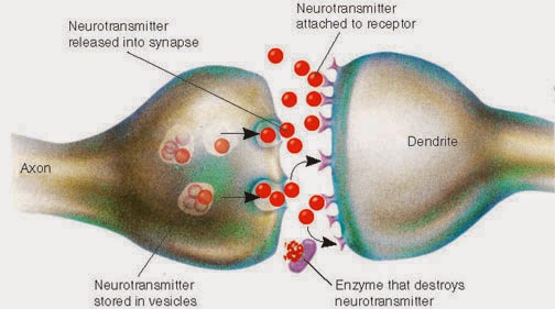
For a little more information and overview of neuron communication, here is a more detailed diagram:

When the electrical signal reaches the end of the axon, a special thing happens with Calcium entering the cell which allows those vesicles full of Neurotransmitter chemicals to exocytose. That's a fancy word meaning the vesicle smooshes into the cell membrane, kicking its contents outside the cell- into the synapse. Those neurotransmitters are then free to act on the postsynaptic cell dendrites or body to continue the message along!
But how did those vesicles of neurotransmitter actually GET there? The cell factories that make neurotransmitters which are mostly made of protein, are waaaaaay back at the cell body, but it has to be released at the terminal button of the axon! Axons can be very long, like so:

Neurotransmitter vesicles are continually made by the neuron (each neuron only makes one particular type of neurotransmitter), and sent down the axon. They get there the same way you would travel a long distance- on a "highway" of sorts.
Here is a great animation of the whole neuron communication process, and you will see vesicles coming down to the end of the axon.
So, the cool thing is those vesicles are literally "walked" down the microtubule highway. In my favorite video, you can see this happen:
Lastly, I found this funny little video about the life of a motor protein (the alien-looking guy that walked the vesicle down the microtubule). Enjoy!
P.S. While searching for images, I ran across this amazing blog, so here is a great reference on the brain and neuroscience, explained in simple terms in much the same way I try to do my own blog. The Brain Geek
Stay curious!
-Julie
Here I will summarize what it takes for a message to be passed from one neuron to another. If you are interested in information about the specific channels that make neuron communication possible, see this post on Neurons.
When an action potential - the electrical signals that neurons use to send messages - reaches the end of an axon, the electrical message must become a chemical message in order to cross the synapse. Neurons are connected to each other by a synapse which consists of the axon of the presynaptic neuron (the one sending the message), and the dendrite or cell body of the postsynaptic neuron (the one receiving the message), and the space between them called the synaptic cleft. Chemicals have to be sent across this space for the message to continue on. The chemicals used are neurotransmitters.
Here's a simple diagram of a synapse:
For a little more information and overview of neuron communication, here is a more detailed diagram:
When the electrical signal reaches the end of the axon, a special thing happens with Calcium entering the cell which allows those vesicles full of Neurotransmitter chemicals to exocytose. That's a fancy word meaning the vesicle smooshes into the cell membrane, kicking its contents outside the cell- into the synapse. Those neurotransmitters are then free to act on the postsynaptic cell dendrites or body to continue the message along!
But how did those vesicles of neurotransmitter actually GET there? The cell factories that make neurotransmitters which are mostly made of protein, are waaaaaay back at the cell body, but it has to be released at the terminal button of the axon! Axons can be very long, like so:
Neurotransmitter vesicles are continually made by the neuron (each neuron only makes one particular type of neurotransmitter), and sent down the axon. They get there the same way you would travel a long distance- on a "highway" of sorts.
Here is a great animation of the whole neuron communication process, and you will see vesicles coming down to the end of the axon.
So, the cool thing is those vesicles are literally "walked" down the microtubule highway. In my favorite video, you can see this happen:
Lastly, I found this funny little video about the life of a motor protein (the alien-looking guy that walked the vesicle down the microtubule). Enjoy!
P.S. While searching for images, I ran across this amazing blog, so here is a great reference on the brain and neuroscience, explained in simple terms in much the same way I try to do my own blog. The Brain Geek
Stay curious!
-Julie
Subscribe to:
Posts (Atom)

