Let's talk about the most amazing organ in the most complex organism on the planet, shall we? :)
Here is a basic orientation:
Brain Stem
The most ancient ("primitive") part of the brain is the brain stem. This part of your brain is responsible for basic functions to keep you alive. Includes the Medulla oblongata, Pons, and Midbrain. First a look at the parts:
Here is a view of a frontal slice through the human brain stem:
 |
| 1- Cerebrum, 2- thalamus, 3-midbrain, 4- pons, 5- medulla oblongata, 6- spinal cord (Source) |
|
|
|
|
|
|
Now a look at each part and the centers each contains that help keep you alive in-the-moment. Things that could kill you in a matter of seconds or minutes if they don't work properly are controlled here. We'll start with the most inferior part and move up.
Medulla Oblongota
Control centers of Medulla:
- Vasomotor- regulate blood pressure
- Cardiac center - controls heart rate, strength of contraction
- Respiratory center - controls respiratory muscles, is sensitive to pH (so it can respond by increasing or decreasing respiratory rate which will bring the body back into homeostasis as far as pH)
- Respiratory reflexes - vomiting, swallowing, coughing, sneezing
Mnemonic from Physiology student - "programming your VCR makes you vomit"
Another cool thing that happens in the medulla:
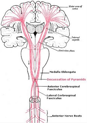
- Decussation of fiber tracts - decussation is a fancy scientific
word for swapping. As you all know, the right side of the brain
controls the left side of the body, and vice verse. The medulla is
where the nerve fibers cross over. (Also referred to as "decussation of pyramids".)
Pons
- Pathway between spinal cord and brain
- Fine control of respiration
 |
| Inferior view (looking up at bottom of brain) showing the Pons (Wikipedia) |
Mesencephalon (midbrain)
- Conduction pathway between cerebral hemispheres
- Reflexes based on visual stimuli (moves head and eyes to protect them from being hit or poked)
That is the brain stem. Next we will continue superiorly.
Thalamus
Paired organ at top of brain stem, see picture below
 |
| Thalamus is the purple part. Detailed anatomy of brain stem if you want that info. (Source) |
- General sensations of pain, heat, cold, pressure (can't pinpoint location or intensity)
- Sensory Gatekeeper - pathway between cerebral cortex and sensory/somatic systems. Shuts out some sensory info to allow for greater focus on important things. So it takes in all that general info and filters out what's not important (such as the sensation of your clothes touching your skin so stuff like that doesn't become distracting)
Reticular Activating System
Housed in the brainstem and thalamus is the Reticular Activating System, shown below in red.
This system controls your waking state and alertness, sending signals to the cerebral cortex to keep you awake and alert. This allows you to direct your attention to specific events. It also holds back sensory info at other times to allow you to sleep, and controls your circadian rhythms (sleep-wake transition). In fact, if I understand correctly, the RAS has an inhibatory affect on the part of the brain that contributes to sleepiness - the Preoptic Area (POA) of the Hypothalamus. Likewise, the POA can inhibit the RAS when it's time to sleep. Pretty cool.
An even more cool fact - this alertness center is very active when awake, and is suppressed during deep sleep, but during REM sleep when you are dreaming, the RAS is just as active as when you are awake! :)
Hypothalamus
The inferior part (floor) of the thalamus. See picture above, the purple front section on left picture. Also, the picture below gives a more general orientation. Here are its functions.
- Manager of the Autonomic Nervous System
- Rage & aggression
- Hunger & satiety
- Fluid osmolarity
- Thirst center
- Anti-diuretic hormone (ADH)
- Body temp
- Regulates the Anterior Pituitary Gland, which affects:
- Growth
- Sexual Function
- Lactation (breast feeding), regulated by oxytocin
- Thyroid function
- Stress, regulated by cortisol
Here's a more detailed diagram of the hypothalamus:
Cerebellum
The motor integrating center - makes last-minute fine tuning adjustments to motor activities, controlling coordination.
Tells "how far, how fast, and when to put the brakes on"
Sobriety tests are really testing the functionality of the cerebellum. Depends on sight and proprioception (info on what your body position actually is right now) input. Prevents dysmetria and past-pointing. (Dysmetria = missing the mark; past-pointing = moving beyond the mark.)
The cerebellum processes input from other areas of the brain, spinal
cord and sensory receptors to provide precise timing for coordinated,
smooth movements of the skeletal muscular system. A stroke affecting the
cerebellum may cause dizziness, nausea, balance and coordination
problems.
Cerebrum
Basal Ganglia/ Basal Nuclei
Trains the brain to do fine motor activities
Suppresses unwanted movement
Initiates trained complex movement patterns
Lesion in this area causes Parkinson's disease
Limbic System
Includes parts surrounding the brain stem: cerebral cortex, basal nuclei, thalamus, and hypothalamus
Responsible for "fight or flight" type of survival, and species survival instincts:
- aggression
- fear
- feeding
- sexual behavior
- motivation
Cortices of the Brain
Primary Motor Cortex
Voluntary contraction of skeletal muscle
Premotor Cortex
Stereotyped movements - "computer programmer" for repeatedly used actions
*Broca's Area
Controls muscles responsible for speech - only on the left side of the brain
Somatosensory Cortex
Pinpoints location and intensity of temperature, touch, pressure, and pain (general sensations of these come from thalamus, then are passed on here to get more details)
Proprioception
Primary Visual Cortex
Vision
Primary Auditory Cortex
Hearing
Olfactory Cortex
Smell
Gustatory Cortex
Taste
Parietal-Temporal-Occipital Association Cortex
Interprets primary sensations from somatic, auditory, and visual areas
*Wernicke's area
Allows for interpretation/ understanding of language (both written and spoken)
Prefrontal Association Cortex
Planning for voluntary activities
Perceiving consequences of future actions
Responsible for personality traits
Teen drinking impairs the development in this area
For additional information related to the brain, see these other posts:
Action Potentials
Action Potentials Up Close
Neurons
Neuroglia
Brain Food: Nutrition for Learning & Memory
Nervous System


