This is super cool! Characterizations of the first 12 elements of the Periodic Table of Elements.
http://kcd-elements.tumblr.com/
Sunday, August 2, 2015


Tuesday, July 14, 2015
Pluto
New Horizons came closest to Pluto today, releasing some amazing images! So sad Pluto was demoted from planet status before we got there, but this is still really exciting, and more pictures will be out tomorrow.
Click here to read more on NASA's website.
I couldn't resist making this meme, since so far it looks like no one else has (which surprised me). What else do you do when you have a planet with what looks like a giant heart on it? hah
Stay curious!
~Bio Geo Nerd
Click here to read more on NASA's website.
I couldn't resist making this meme, since so far it looks like no one else has (which surprised me). What else do you do when you have a planet with what looks like a giant heart on it? hah
 |
| I love you to Pluto and back |
Stay curious!
~Bio Geo Nerd
Sunday, May 17, 2015
Marie Tharp - Geologist

Marie Tharp was responsible for verifying Alfred Wegener's theory of Continental Drift, a key Theory in Geology. Read all about her in this amazing article on mentalfloss "How One Woman's Discovery Shook the Foundations of Geology".
Wegener's was a theory the scientific community only laughed at when presented in 1912 until all the evidence came pouring in and staring them in the face many years after Wegener's death. The theory was laughed at, and according to Marie Tharp's recollection, you could even be fired from geologic work for believing his theory.
As an aside, here's a fun video on Wegener
Even when Marie Tharp first showed the evidence - a map she had made from radar data of the sea floor which clearly showed the mid-ocean ridge where sea floor spreading takes place and allows for continental drift - to her colleague in the 1950's, he made her redo all her hard work mapping the seafloor because he couldn't believe it backed up Wegener's Theory and pegged it as only "girl talk". She started from scratch and came up with the same thing, which also was corroborated by this colleague's own data of undersea earthquakes.
This laid the way for Harry Hess to peg Seafloor Spreading as the mechanism for Continental Drift. Wegener and Hess are recognized as the fathers of Plate Tectonics in modern Geology.
I'm going to go out on a limb and consider Tharp as a mother of Plate Tectonics! The only reason she wasn't out on research ships collecting radar data herself for this work was because they wouldn't allow women on ships- it was considered bad luck. In fact the only way she even got to study geology was because after Pearl Harbor, universities finally opened up their programs to women. Marie Tharp is not well known, but she managed to triumph over the sexism of the time and is now one of my heroes.
References
This post was spurred by this amazing article, which goes into detail about what Marie Tharp did. Do yourself a favor and read it! (Picture also from there)
http://mentalfloss.com/article/60481/how-one-womans-discovery-shook-foundations-geology
http://en.wikipedia.org/wiki/Alfred_Wegener
Wednesday, March 4, 2015
The Teen Brain
Can I just tell you how much I love teenagers? I am student teaching at a high school and I thoroughly enjoy being around these young people, with their varied personalities, interests, challenges, goals, etc.. I obviously adore science, but these kids make my job so great I'd probably still like it even if I didn't get to teach about my favorite thing ever. I just wish they all knew how awesome they are and can be.
Adolescence is a really interesting conundrum in the development of a human being. It's an exciting time that leads up to so many big life changes- getting through school, choosing a career, starting a family, making a place for oneself in the world. So many physical changes are happening, not the least of which are the changes in the brain. These changes make it both easier and more difficult for people to become responsible adults, which is one reason it is so incredibly fascinating, and makes me truly appreciate and empathize with teenagers that much more.
An explosion of brain development
It is well-known that humans go through an explosion of brain development during the first 3 years of life. Infants and toddlers are actively exploring their worlds and making many connections in their brains. But did you know this explosion also happens in adolescence? And interestingly, an important part of this development is cutting away parts that are no longer needed. This process is called "synaptic pruning".
Synaptic Pruning
This term refers to the "synapses" which are the connections between brain cells. Cutting those away may seem counter-intuitive, but it is actually a wonderful part of developing a faster, more efficient brain. It's a very exciting time for learning.
Here are some photographs taken through a microscope of brain cells at various ages. Notice how there is an explosion of growth as the baby develops, and then as they become school-aged, things are pruned away while what's left becomes strengthened.

I have put in a couple of great video clips to explain this process.
Short, student-friendly explanation:
More detailed (fascinating!) info on synaptic pruning - TED talk by Sarah-Jayne Blakemore:
Prefrontal Cortex Development
If you watched that second video above, you heard a lot about how the adolescent prefrontal cortex is developing. This area of the brain is very important for higher-order or "executive" functioning skills. That includes things like planning, predicting consequences, impulse control, and personality.

So the interesting thing is.... this area of the brain is not fully developed in humans until around age 25! Does the sometimes irrational behavior of teenagers make more sense given that information? Teens are very prone to risk-taking, which makes sense given the fact that they are still learning how to control impulses, predict consequences, and make good decisions. This lower impulse control can also be an amazingly good thing! So many people have made amazing discoveries, inventions, etc. while in this stage of brain development, because they had all the drive to go for their goal, without their brains putting the brakes on. So while impulse control is super important, sometimes it can be overdone and stifle creativity. Without that fully in place, teens can do some amazing things - amazingly stupid or amazingly creative and exceptional!
Societal influences
So the conundrum I mentioned is that a teenager has this amazing brain with abilities and creativity, but heightened propensity toward risk taking and foolishness and less ability to control those impulses. And yet this is a time period when people are expected to make really important decisions that will shape the rest of their lives.
It is also a time when many young people are filling those moldable brains of theirs with garbage! Not just garbage that will prove pretty useless in the future, like Angry Birds and Candy Crush. But garbage that is going to forever stunt their progress and could even ruin their lives. Alcohol, pornography, and drugs can wreak havoc on a developing brain and the person whose head it's in. But those are the things that are exciting and new, and the teen hasn't yet developed full ability to put the brakes on.
That, however is NOT an excuse. It is important for teenagers to realize the limitations of being a teenager and rely on advice of parents, guardians, teachers, and other trusted adults to help keep them safe. It makes perfect sense that we strive to educate young people about staying away from risky things like drugs before they even get to be teenagers. If a young person can decide while they are a child that they will say no to drugs, no questions asked...that choice has already been made and they won't have to grapple as hard with it later on. They will also be helped by the added protection of choosing good friends who have also made similar choices for themselves.
So to wrap this up.... teenagers are awesome creatures. They have a LOT of brain work to do, while working with a less developed instrument than those of adults. They need good adults who will support and guide them. Do what you can to help educate them, but above all, love them and cut them a little slack.
Adolescence is a really interesting conundrum in the development of a human being. It's an exciting time that leads up to so many big life changes- getting through school, choosing a career, starting a family, making a place for oneself in the world. So many physical changes are happening, not the least of which are the changes in the brain. These changes make it both easier and more difficult for people to become responsible adults, which is one reason it is so incredibly fascinating, and makes me truly appreciate and empathize with teenagers that much more.
An explosion of brain development
It is well-known that humans go through an explosion of brain development during the first 3 years of life. Infants and toddlers are actively exploring their worlds and making many connections in their brains. But did you know this explosion also happens in adolescence? And interestingly, an important part of this development is cutting away parts that are no longer needed. This process is called "synaptic pruning".
Synaptic Pruning
This term refers to the "synapses" which are the connections between brain cells. Cutting those away may seem counter-intuitive, but it is actually a wonderful part of developing a faster, more efficient brain. It's a very exciting time for learning.
Here are some photographs taken through a microscope of brain cells at various ages. Notice how there is an explosion of growth as the baby develops, and then as they become school-aged, things are pruned away while what's left becomes strengthened.
I have put in a couple of great video clips to explain this process.
Short, student-friendly explanation:
More detailed (fascinating!) info on synaptic pruning - TED talk by Sarah-Jayne Blakemore:
Prefrontal Cortex Development
If you watched that second video above, you heard a lot about how the adolescent prefrontal cortex is developing. This area of the brain is very important for higher-order or "executive" functioning skills. That includes things like planning, predicting consequences, impulse control, and personality.
So the interesting thing is.... this area of the brain is not fully developed in humans until around age 25! Does the sometimes irrational behavior of teenagers make more sense given that information? Teens are very prone to risk-taking, which makes sense given the fact that they are still learning how to control impulses, predict consequences, and make good decisions. This lower impulse control can also be an amazingly good thing! So many people have made amazing discoveries, inventions, etc. while in this stage of brain development, because they had all the drive to go for their goal, without their brains putting the brakes on. So while impulse control is super important, sometimes it can be overdone and stifle creativity. Without that fully in place, teens can do some amazing things - amazingly stupid or amazingly creative and exceptional!
Societal influences
So the conundrum I mentioned is that a teenager has this amazing brain with abilities and creativity, but heightened propensity toward risk taking and foolishness and less ability to control those impulses. And yet this is a time period when people are expected to make really important decisions that will shape the rest of their lives.
It is also a time when many young people are filling those moldable brains of theirs with garbage! Not just garbage that will prove pretty useless in the future, like Angry Birds and Candy Crush. But garbage that is going to forever stunt their progress and could even ruin their lives. Alcohol, pornography, and drugs can wreak havoc on a developing brain and the person whose head it's in. But those are the things that are exciting and new, and the teen hasn't yet developed full ability to put the brakes on.
That, however is NOT an excuse. It is important for teenagers to realize the limitations of being a teenager and rely on advice of parents, guardians, teachers, and other trusted adults to help keep them safe. It makes perfect sense that we strive to educate young people about staying away from risky things like drugs before they even get to be teenagers. If a young person can decide while they are a child that they will say no to drugs, no questions asked...that choice has already been made and they won't have to grapple as hard with it later on. They will also be helped by the added protection of choosing good friends who have also made similar choices for themselves.
So to wrap this up.... teenagers are awesome creatures. They have a LOT of brain work to do, while working with a less developed instrument than those of adults. They need good adults who will support and guide them. Do what you can to help educate them, but above all, love them and cut them a little slack.
Thursday, January 1, 2015
Your Metabolism Doesn't Know It's The New Year
This is a more scientific, detailed continuation of a discussion started on my other blog. Fat Fun Fit: Why I'm Not Making Resolutions: A Case Against Dieting. From that post (in blue):
Continuing on....
Your brain needs glucose. Your body does too. It's the energy that gets broken down into usable energy for your body, known as ATP. You can also use other simple sugars like fructose, but those actually just get converted to glucose before being used to make energy.
Glucose can be stored in a number of ways. The easiest way is as glycogen. That is a ginormous molecule of glucoses linked together in a network. Your liver and muscles store glycogen so that your body and brain have enough energy between meals. They can only store enough to last for a few hours (between meals). When there is no glucose in your blood from a recent meal and there is a need for energy, the glycogen gets broken down to glucose.
In the muscle glycogen, the glucose is used right there in the muscles so you can still walk around, type at your computer, chase your kids, etc. between meals. The liver is the bank account for your brain. The glycogen in the liver is broken down to send glucose into the blood for the brain to use. Brain always has priority. If the brain doesn't get enough, the body must supply.
When glycogen is gone, the body will go to the next source. Muscle. There are a lot of Amino Acids - building blocks of proteins - in your muscles, cause that's what muscles are made of- loads and loads of proteins. Those proteins can then be made back into glucose to send to the brain so it has energy. This process is called gluconeogenesis (gluco=glucose, neo= new, genesis= make; make new glucose).
When you are on a very low-calorie diet or starvation, your body will break down some muscle to feed your brain. In the absence of readily useable glucose in the diet, the same happens. This is why people on Atkins/ low carb diets lose a lot of weight quickly at the beginning. They are starving their brains of glucose, so the muscle gets broken down. Well, muscle weighs A LOT. It is much more dense and heavy than fat. It also takes a lot more water to metabolize muscle, so the majority of those pounds you are dropping on those diets are muscle and water. Great if all you care about is the number on the scale. Terrible if you care about your body composition, shape/ size, and actual health.
The other way for glucose to be stored is for PARTS of it to be put into fat. Glucose has 6 Carbons in it, but a bunch of 2-carbon subunits (Acetyl-CoA) can be put together into a long chain: a fatty acid. Then 3 of these chains can be attached to a glycerol and stuck in adipocytes- fat cells. A lot of energy can be tucked away this way. You get loads and loads of energy out of fat. You are probably familiar with this if you pay attention to nutrition labels. There are 9 Calories per gram of fat, but only 4 Calories per gram of carbohydrate or protein. That's equalizing the weight. Fat takes up a lot more space, so all that extra energy stored in your body also makes your body bigger than the same amount of energy stored in muscles or glycogen.
So, how and when does this fat energy get used? That's the tricky thing. Your brain lacks the proper gateways and enzymes to metabolize the subunits from fatty acids (acetyl CoA) directly. It can only take in glucose or ketone bodies. The glycerol from the triglyceride can be made into glucose, so those get sent off to the liver to do that. But the fatty acids are better used by the cells of the body cause they have the right enzymes and gates to allow that to happen.
But since fat is long-term storage, just like a trust fund, the body is not going to break into it at the first sign of trouble. It waits to see if it can get by with the cash on hand, the checking and savings account. (Glucose in blood from your meal, then glycogen, then muscle as explained already.) Then if it's dire enough it will go for those fatty acids IF the body is in need of it. So, from what I have heard from a Physiology professor about some research (but haven't located the research myself yet so I have no link, sorry), is that the mark for the fat getting mobilized is about 45 minutes of exercise. After that point you would start to break down the fat for your body to use. So the recommendation of 30 minutes of exercise most days doesn't even touch that. If I want to reduce body fat, the best thing is to go on hours-long hikes as much as possible. (The regular aerobic exercise IS crucial to your health in other ways though, and should still be maintained for your cardiovascular, respiratory, muscle and mental health. Benefits of regular exercise are nearly endless.)
I mentioned the brain can use ketone bodies. In severe starvation, the fatty acids can be made into ketone bodies, which can get into the brain and used for energy. But they are very dangerous because they turn the blood acidic and it is detrimental to your body. This is the last ditch effort during severe starvation, to keep your brain alive, cause without your brain, the show is over.
How will your body react if your norm is to eat around 2,500 calories
per day, and only a little walking as your physical activity, and you
suddenly cut your food intake by HALF and jump up to a "perfect" workout
regimen? My body reacted like this, as would most: "Holy crud!! What
the hell is going on?!! We must be starving, and running to try to
find food! What apocalypse is going on out there? Oh man, this is it,
this is the end. Emergency! Emergency! Going to code red- crisis
management mode!" My body senses a sudden catastrophic world event on
many January 1st's.
Now time for the science nerd in
me to come out. What happens physiologically while on a low-calorie
diet that your body is not used to, is that all food coming in as much
as possible will be stored as fat for later in case the emergency gets
even worse, and muscle gets broken down to be used for quick energy.
Cause if you're in a crisis, keeping your brain well-fed, and planning
for the worst is top priority. Your brain is not able to use energy
from fat, and if you are starving your brain by eating a severely
low-calorie diet, muscle is the quickest way to get energy to your
brain. Your body doesn't really have a way to tell how much fat it
already has stored so that it can "cap" it at a certain amount, which is
why you can get very morbidly obese people. Your body will just keep
making more fat when it is in these crises even though you already have a
lot, or when there is a plain old excess of energy coming in. Losing that muscle lowers my energy and makes sustaining this plan much more difficult.
Continuing on....
Your brain needs glucose. Your body does too. It's the energy that gets broken down into usable energy for your body, known as ATP. You can also use other simple sugars like fructose, but those actually just get converted to glucose before being used to make energy.
Glucose can be stored in a number of ways. The easiest way is as glycogen. That is a ginormous molecule of glucoses linked together in a network. Your liver and muscles store glycogen so that your body and brain have enough energy between meals. They can only store enough to last for a few hours (between meals). When there is no glucose in your blood from a recent meal and there is a need for energy, the glycogen gets broken down to glucose.
In the muscle glycogen, the glucose is used right there in the muscles so you can still walk around, type at your computer, chase your kids, etc. between meals. The liver is the bank account for your brain. The glycogen in the liver is broken down to send glucose into the blood for the brain to use. Brain always has priority. If the brain doesn't get enough, the body must supply.
When glycogen is gone, the body will go to the next source. Muscle. There are a lot of Amino Acids - building blocks of proteins - in your muscles, cause that's what muscles are made of- loads and loads of proteins. Those proteins can then be made back into glucose to send to the brain so it has energy. This process is called gluconeogenesis (gluco=glucose, neo= new, genesis= make; make new glucose).
When you are on a very low-calorie diet or starvation, your body will break down some muscle to feed your brain. In the absence of readily useable glucose in the diet, the same happens. This is why people on Atkins/ low carb diets lose a lot of weight quickly at the beginning. They are starving their brains of glucose, so the muscle gets broken down. Well, muscle weighs A LOT. It is much more dense and heavy than fat. It also takes a lot more water to metabolize muscle, so the majority of those pounds you are dropping on those diets are muscle and water. Great if all you care about is the number on the scale. Terrible if you care about your body composition, shape/ size, and actual health.
The other way for glucose to be stored is for PARTS of it to be put into fat. Glucose has 6 Carbons in it, but a bunch of 2-carbon subunits (Acetyl-CoA) can be put together into a long chain: a fatty acid. Then 3 of these chains can be attached to a glycerol and stuck in adipocytes- fat cells. A lot of energy can be tucked away this way. You get loads and loads of energy out of fat. You are probably familiar with this if you pay attention to nutrition labels. There are 9 Calories per gram of fat, but only 4 Calories per gram of carbohydrate or protein. That's equalizing the weight. Fat takes up a lot more space, so all that extra energy stored in your body also makes your body bigger than the same amount of energy stored in muscles or glycogen.
So, how and when does this fat energy get used? That's the tricky thing. Your brain lacks the proper gateways and enzymes to metabolize the subunits from fatty acids (acetyl CoA) directly. It can only take in glucose or ketone bodies. The glycerol from the triglyceride can be made into glucose, so those get sent off to the liver to do that. But the fatty acids are better used by the cells of the body cause they have the right enzymes and gates to allow that to happen.
But since fat is long-term storage, just like a trust fund, the body is not going to break into it at the first sign of trouble. It waits to see if it can get by with the cash on hand, the checking and savings account. (Glucose in blood from your meal, then glycogen, then muscle as explained already.) Then if it's dire enough it will go for those fatty acids IF the body is in need of it. So, from what I have heard from a Physiology professor about some research (but haven't located the research myself yet so I have no link, sorry), is that the mark for the fat getting mobilized is about 45 minutes of exercise. After that point you would start to break down the fat for your body to use. So the recommendation of 30 minutes of exercise most days doesn't even touch that. If I want to reduce body fat, the best thing is to go on hours-long hikes as much as possible. (The regular aerobic exercise IS crucial to your health in other ways though, and should still be maintained for your cardiovascular, respiratory, muscle and mental health. Benefits of regular exercise are nearly endless.)
I mentioned the brain can use ketone bodies. In severe starvation, the fatty acids can be made into ketone bodies, which can get into the brain and used for energy. But they are very dangerous because they turn the blood acidic and it is detrimental to your body. This is the last ditch effort during severe starvation, to keep your brain alive, cause without your brain, the show is over.
Thursday, October 30, 2014
Memory & Amnesia
(This post has resources and information for learning about Memory and Amnesia, for Physiological Psychology.)
Radio Lab:
6:30 Rat tests, 8:08 Chemical prevent memory in rats; Clive Waring 42:30
The Hippocampus and Patient H.M.(by Ted Ed)
H.M. - Nova special
9:22 What like for H.M. 10:40-11:52 Muscle memory star test
Morris Water Maze
Radio Lab:
6:30 Rat tests, 8:08 Chemical prevent memory in rats; Clive Waring 42:30
The Hippocampus and Patient H.M.(by Ted Ed)
H.M. - Nova special
9:22 What like for H.M. 10:40-11:52 Muscle memory star test
Morris Water Maze
Friday, October 24, 2014
Periodic Table of Cookies
In honor of chemistry week,
Here's the Periodic Table of Cookies my club (Science Association of Women at UVU) did this spring to make money. Making that many cookies was insane. But fun. :)


Here's the Periodic Table of Cookies my club (Science Association of Women at UVU) did this spring to make money. Making that many cookies was insane. But fun. :)
 |
| Printed on sugar overlay, we cut each square out |
Tuesday, October 21, 2014
Footprints and trackways
How do paleontologists and anthropologists use footprints and trackways? Often, information is extrapolated from tracks and used to determine approximate heights and sizes of the organism that left the tracks. How can this be? Is there a correlation between foot size, leg length, stride length...and HEIGHT? Let's find out!
.JPG)

.JPG)

.JPG)


These formulae don't only work for dinosaurs. If you like Lego, you may like to know that using Alexander and Thulborn's formulae, a Lego minifig (the little people) could walk at 0.5 km/h and run at 2.2 km/h
(Lego info from http://www.csiro.au/helix/sciencemail/activities/dinospeed.html)
![Speed (in metres per second)=0.25 x (square root of g) x [stride length]^1.67 x [hip height]^-1.17](https://lh3.googleusercontent.com/blogger_img_proxy/AEn0k_snlYewb6B9b1gUJOLpIzefZRvRLA2Ja_Wv6EOQMLHT9ApN2qvGlyM6bHmHDFZLgBwmaKoiFPLkS8i-js-uLHeTbD1CUiwuGBeDXoW9Pbkzn9Sjv7el7pKc_U1J2TH_oS6X2YcTO01KBpiPpNA=s0-d)
Where g is the acceleration from gravity. On Earth, g=9.8 m/s². Don't worry if you haven't come across maths like this before. What's important is that it's a way of estimating an animal's speed, without measuring it directly.
![Speed (in metres per second)=7.8 x ( 200/[number of steps] )^1.67 x [hip height]^-1.17](https://lh3.googleusercontent.com/blogger_img_proxy/AEn0k_u2nFrOBuQqQUfi0gQocS1e8ZZMpULoWbrKXYG5L70BAEX4cPcKGRXJ70E_LhD7X_Flz2qCMPUfeQl7SSVJawL0CRSejatBDL_rYvBae5Df4IpEyibMtpNtCEsoVL66HI8CzJDTSdD95d38wKkO=s0-d) Your actual speed is just the distance divided by the time it took:
Your actual speed is just the distance divided by the time it took:

The basic measurement of a dinosaur footprint is its length, represented as FL. The ratio of footprint length and hip height (h) is different for different groups of dinosaurs, but generally the hip height of a bipedal dinosaur is roughly four times the footprint length. The speed can then be determined as relative speed, which is stride length (SL), divided by hip height (h). Generally speaking, if the SL/h <2.0, then the animal was walking; >2.9, the animal was running; and between 2.0 and 2.9, the animal was trotting.1 (Source: http://www.ucmp.berkeley.edu/education/dynamic/session3/sess3_act2.htm)
These formulae don't only work for dinosaurs. If you like Lego, you may like to know that using Alexander and Thulborn's formulae, a Lego minifig (the little people) could walk at 0.5 km/h and run at 2.2 km/h
 |
| Using the same equations that estimate dinosaurs' speeds, Lego figures could run at 2 km/h and walk at 0.5 |
![Speed (in metres per second)=0.25 x (square root of g) x [stride length]^1.67 x [hip height]^-1.17](http://www.csiro.au/helix/sciencemail/activities/images/DinoAlexander1.gif)
Where g is the acceleration from gravity. On Earth, g=9.8 m/s². Don't worry if you haven't come across maths like this before. What's important is that it's a way of estimating an animal's speed, without measuring it directly.
We can check Alexander's formula
by comparing it to our measurements. The stride length is just the
distance (100m) divided by the number of strides (half the number of
steps). In terms of your measurements, Alexander's formula becomes:
The basic measurement of a dinosaur footprint is its length, represented as FL. The ratio of footprint length and hip height (h) is different for different groups of dinosaurs, but generally the hip height of a bipedal dinosaur is roughly four times the footprint length. The speed can then be determined as relative speed, which is stride length (SL), divided by hip height (h). Generally speaking, if the SL/h <2.0, then the animal was walking; >2.9, the animal was running; and between 2.0 and 2.9, the animal was trotting.1 (Source: http://www.ucmp.berkeley.edu/education/dynamic/session3/sess3_act2.htm)
What can a single track tell us?
Not too much. We can infer something about the size of the animal and maybe something about the sediment. We may have a clue as to who made the track.What can a trackway (a series of tracks or footprints) tell you?
-
Who was there.
-
How many animals were there.
-
The sizes of the animals compared to one another.
-
How they were interacting - social activity, such as herds,
moving in families, etc.
-
How fast they were moving.
-
What the sediment was like, and therefore something about the
environment of the time.
Source of above text: http://www.ucmp.berkeley.edu/education/dynamic/session3/sess3_stories3.htm
Thursday, October 2, 2014
Leaf Anatomy
View of a shoot apical meristem with some leaf primordia. Additionally, I have labelled the 3 primary meristems you can differentiate here.

Here's a nicotine leaf to show the two kinds of meristems specific to the leaf. The leaf apical meristem becomes the midrib of the leaf, while the leaf marginal meristem is the blade of the leaf.

Epidermis
From a Sedum (stone crop)- you can see the "regular" epidermal cells (squiggly shaped) as well as the guard cells in various places surrounding stomata.

Take a closer look:

Also here is a sunken stomate as seen in a Pinus leaf. It is "sunken" because the guard cells are below the level of the epidermis which helps protect against dessication (drying out).

Mesophyll
Mesophyll just means middle of the leaf. It is the term for the ground tissue in the leaf. There are two types by shape: palisade and spongy.
You can see both of these well in a pine leaf:

We also term a leaf to be unifacial or bifacial based on the arrangement of the mesophyll. Bifacial is if there is spongy on one side, and palisade on the other. Unifacial is either the same type throughout, or it makes a kind of sandwich with the same kinds on either side and something different in the middle. You can see this type of unifacial in a Dianthus (carnation) leaf:

Pine anatomy
Some things we learned specifically with pine needles (I'm not sure if they also apply to some other plants, sorry) are the resin duct with epithelium, hypodermal sclerenchyma, and transfusion tissue.
Some of these are labeled:

The outermost layer surrounding the resin duct is the epithelium. Hypodermal sclerenchyma is below ("hypo") the outermost layer of cells or dermis. These have thick secondary cell walls which add to the strength of a pine needle, as you have probably tested yourself many times when you got poked with one. In this repeat picture you can see the hypodermal sclerenchyma with the thick red-stained walls, on either side of the sunken stomate.
Bundle Sheaths
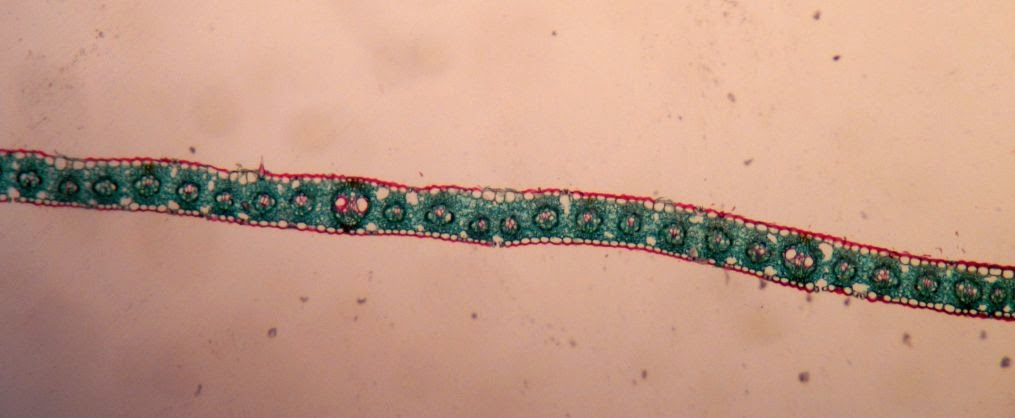
Bundle sheaths are different in C3 plants and C4 plants.
Here, in a C3 grass (Poa), you see their regular bundle sheaths:

Closer. Just looks like a blank set of cells surrounding the vascular bundle.

But in a C4 plant, like this Zea corn, the sheaths have what is referred to as "Kranz anatomy". Kranz is German for wreath, and you can see they are rather leaf-like in the following examples. There are two orientation pictures, then it zooms in one a single bundle sheath so you can see the Kranz anatomy.


Leaf Abscission Zone
When a plant loses its leaves, it prepares for this by creating an abscission zone so the leaf can easily fall off without damaging any of the other tissue. Layers of cells secrete suberin (or is it subirin?) for protection (and are called suberized cells), while the next outermost layer of cells is pre-programmed to break easily, as it were. The weak layer is called the separation layer.

Whew, that's a lot of leaf anatomy! Stay curious.
P.S. There are no picture source references cause I took all these with my camera (through the microscope in Botany lab) and did the labeling myself.
Tuesday, September 9, 2014
Plant Cell Wall Lab
In this lab we explored various anatomical features of plant cell walls.
Mitosis & Cytokinesis
Not going to repost that, I did this last year and it is still beautiful: Mitosis post
Here's another great pic of a cell plate and phragmoplast on the edges I got today though:

Cytokinesis in plants is a rather involved process since a new middle lamella must be made, followed by primary wall and sometimes secondary wall.
Primary pit fields
This is a TERRIBLE picture taken of a picture shown to me on another camera. But you can kind of see the primary wall and the thinner parts that indicate the primary pit fields (pointer is on the thicker part of the primary cell wall).

More primary pit fields from a more 3D outside view of the cell (this is from #6 of the lab).

The cell walls are stained pink and you can see that there are lighter/ white spots on it. That's where the wall is thinner, so those are the primary pit fields.
Plasmodesmata
By definition, plasmodesmata are only in primary cell walls, and they are channels between adjacent cells, through which the cytoplasm of each cell is continuous. These occur more often in primary pit fields where the membrane is thinner, but they can happen anywhere in the primary wall.
 You can also see these in the tomato cells we looked at last week:

Intercellular Air Spaces
These get formed often at the edges of cells as they are dividing, and are important to gas exchange for the plant. (#6)

Secondary Cell Wall
Here you can see the distinction between the secondary wall and the compound middle lamella (which includes middle lamella and 2 adjacent primary walls).
 The lighter lines between all the cells are the compound middle lamella, the darker parts are secondary cell wall.
Pits
These are pear stone cells, and they have many long simple pits running through their secondary cell walls. I thought they look rather similar to plasmodesmata, but the distinction is plasmodesmata are ONLY in primary walls, and pits are ONLY in secondary walls. (Lab #9)

Bordered pits
Bordered pits from above look like little donuts. Here there are a whole bunch in some dense pine wood. These types of pits are common in water conducting cells, and they act to help prevent clogs from air bubbles. (Lab #11)


Stained microscope slides are pretty. That is all.
Stay curious.
Mitosis & Cytokinesis
Not going to repost that, I did this last year and it is still beautiful: Mitosis post
Here's another great pic of a cell plate and phragmoplast on the edges I got today though:
Cytokinesis in plants is a rather involved process since a new middle lamella must be made, followed by primary wall and sometimes secondary wall.
Primary pit fields
This is a TERRIBLE picture taken of a picture shown to me on another camera. But you can kind of see the primary wall and the thinner parts that indicate the primary pit fields (pointer is on the thicker part of the primary cell wall).
More primary pit fields from a more 3D outside view of the cell (this is from #6 of the lab).
The cell walls are stained pink and you can see that there are lighter/ white spots on it. That's where the wall is thinner, so those are the primary pit fields.
Plasmodesmata
By definition, plasmodesmata are only in primary cell walls, and they are channels between adjacent cells, through which the cytoplasm of each cell is continuous. These occur more often in primary pit fields where the membrane is thinner, but they can happen anywhere in the primary wall.
Intercellular Air Spaces
These get formed often at the edges of cells as they are dividing, and are important to gas exchange for the plant. (#6)
Secondary Cell Wall
Here you can see the distinction between the secondary wall and the compound middle lamella (which includes middle lamella and 2 adjacent primary walls).
 |
| Just a picture to orient- vascular bundle, and the bundle cap at the top. |
Pits
These are pear stone cells, and they have many long simple pits running through their secondary cell walls. I thought they look rather similar to plasmodesmata, but the distinction is plasmodesmata are ONLY in primary walls, and pits are ONLY in secondary walls. (Lab #9)
Bordered pits
Bordered pits from above look like little donuts. Here there are a whole bunch in some dense pine wood. These types of pits are common in water conducting cells, and they act to help prevent clogs from air bubbles. (Lab #11)
Stained microscope slides are pretty. That is all.
Stay curious.
Sunday, September 7, 2014
Plant Cell Wall Synthesis
Plants have some things animals don't, including a cell wall surrounding their cells.
Primary Cell Wall
Here's a diagram of the primary cell wall, along with the middle lamella that lies between adjacent plant cells with their respective primary cell walls. Also the regular-old plasma membrane lies internally to all that.
The middle lamella is made of pectins which are the perfect sticky thing to attach a primary wall made of cellulose microfibrils to! The primary wall also has some other stuff to hold it together in a nice meshy business.
Synthesis of Primary Cell Well
This is the coolest part...
When a cell splits and becomes two cells, a new cell wall must be built between them. I'll go into the details of the cytokinesis itself in another post, but after that is done, all that is there is is a middle lamella (again, made of pectins), and a plasma membrane on either side. How does the primary wall end up BETWEEN the plasma mebrane and the middle lamella?!?
Cellulose synthase, that's how. And it's brilliant. In the plasma membrane, there is a complex of proteins embedded that make cellulose. They look like little rosettes, like the ones depicted in blue, below:

The cellulose microfibrils get put together and come out of the external end of the rosettes (closer to the middle lamella). The long cellulose molecules that strengthen the primary wall adhere to the middle lamella, add some cross-linking stuff (pectin, glycans) and there you have it.
The cool part is those rosettes actually move through the plasma membrane, (like wading through mud) guided by microtubules which are on the internal side of the plasma membrane, leaving the trail of cellulose as it goes.
Here are some other diagrams of how this works.
In this one, the blue arrows indicate the direction the rosettes are "wading" through the plasma membrane, "walking" along the orange microtubules beneath.

This shows how the glucose subunits come in from the cytoplasm (purple circles) and are put together into the complex polymer of cellulose. Again, the arrow shows the rosette is moving to the left, leaving a trail of cellulose to the right.
And here we see the yellow plasma membrane cut away partly so we can see the rosettes that pass through and synthesize the microfibrils of cellulose. Each section of the rosette is an enzyme in its own right that puts together the long chains from glucose, which is then wound together into larger and larger units.
Here's what the structure of cellulose looks like broken down, so you can see it's a complex, tightly packed polymer.
The strands of cellulose are arranged pretty randomly in a primary cell wall. The cellulose synthases don't move very quickly, so the cellulose that is spit out goes around rather randomly, much like squeezing a bunch of toothpaste out of a tube- it goes every which way.
Secondary Cell Wall Synthesis
The secondary cell wall is lain down internally to the primary cell wall. It is thick and has 3 layers that are put down one at a time. Each layer has all its cellulose going parallel to each other. But the layers each have different directions/ orientations than one another, as seen in the bottom part of this diagram:
This provides a lot of extra strength, because it is protecting against compression, stretching, tension, etc. in all directions once you have all 3 layers put down. When the cellulose rosettes are laying down cellulose for a secondary cell wall layer, they move more quickly and in regular, straight lines.
That's all she (I) wrote. Stay curious!
 |
| Source of picture |
Primary Cell Wall
Here's a diagram of the primary cell wall, along with the middle lamella that lies between adjacent plant cells with their respective primary cell walls. Also the regular-old plasma membrane lies internally to all that.
 |
| Link to source |
Synthesis of Primary Cell Well
This is the coolest part...
When a cell splits and becomes two cells, a new cell wall must be built between them. I'll go into the details of the cytokinesis itself in another post, but after that is done, all that is there is is a middle lamella (again, made of pectins), and a plasma membrane on either side. How does the primary wall end up BETWEEN the plasma mebrane and the middle lamella?!?
Cellulose synthase, that's how. And it's brilliant. In the plasma membrane, there is a complex of proteins embedded that make cellulose. They look like little rosettes, like the ones depicted in blue, below:
The cool part is those rosettes actually move through the plasma membrane, (like wading through mud) guided by microtubules which are on the internal side of the plasma membrane, leaving the trail of cellulose as it goes.
Here are some other diagrams of how this works.
In this one, the blue arrows indicate the direction the rosettes are "wading" through the plasma membrane, "walking" along the orange microtubules beneath.
This shows how the glucose subunits come in from the cytoplasm (purple circles) and are put together into the complex polymer of cellulose. Again, the arrow shows the rosette is moving to the left, leaving a trail of cellulose to the right.
 |
| Source of image |
And here we see the yellow plasma membrane cut away partly so we can see the rosettes that pass through and synthesize the microfibrils of cellulose. Each section of the rosette is an enzyme in its own right that puts together the long chains from glucose, which is then wound together into larger and larger units.
 |
| Source of image |
Here's what the structure of cellulose looks like broken down, so you can see it's a complex, tightly packed polymer.
 |
| Source of image |
The strands of cellulose are arranged pretty randomly in a primary cell wall. The cellulose synthases don't move very quickly, so the cellulose that is spit out goes around rather randomly, much like squeezing a bunch of toothpaste out of a tube- it goes every which way.
Secondary Cell Wall Synthesis
The secondary cell wall is lain down internally to the primary cell wall. It is thick and has 3 layers that are put down one at a time. Each layer has all its cellulose going parallel to each other. But the layers each have different directions/ orientations than one another, as seen in the bottom part of this diagram:
 |
| Source of image |
This provides a lot of extra strength, because it is protecting against compression, stretching, tension, etc. in all directions once you have all 3 layers put down. When the cellulose rosettes are laying down cellulose for a secondary cell wall layer, they move more quickly and in regular, straight lines.
That's all she (I) wrote. Stay curious!
Subscribe to:
Posts (Atom)
Subscribe To Feed


 Posts
All Comments
Posts
All Comments