Saturday, November 16, 2013

Neuron Ion Channels - Detailed

Inward rectifying potassium channels.  This is what started all this.  Just a little paranthetical aside in my Neuroscience teacher's notes that left me curious.  One thing led to another, and here we are.  Ion channels.  Hope you learn something like I did.  Enjoy!

A plethora of potassium channels
Here is a short video showing the molecular structure of a Potassium channel and how it can be perfectly selective to allow Potassium and not Sodium through.

And look at this beautiful top view of a potassium channel with a little purple potassium ion in the center.


Inward-Rectifying Potassium Channels
What does inward-rectifying mean?  Simply put, it just means that voltage travels inward more easily than outward.  So this name tells us that Potassium will move into a cell when open.
(Example - cardiac muscle cells- responsible for the long refractory period between beats, to avoid tetany; kidneys, regulating potassium ions

Action Potentials: Delayed Rectifier Potassium Channels & A Type Channels (outwardly rectifying)


Tandem Pore Domain Potassium Channels (Leak Channels)

Voltage Gated Potassium Channels


********This post under construction!  I just found a textbook with an entire chapter on ion channels, so after I study for my test I will dive into this and finish the post...  thank you for not hating me too much for leaving you hanging.  You'll just have to subscribe to learn more! :)

Wednesday, October 9, 2013

Marie Curie

This was shared on Facebook by Science is Awesome.


Marie Curie was also denied a seat on the French Academy of Sciences in 1911.  She responded to this by diving into her work and earning her second Nobel Prize a year later for her work with radioactivity.  (There is a measurement of the effects of radiation named after her, the Curie, abbreviated Ci.)  Go to this source for a full article about this story.

Here's a real photograph of her.

You can read a short biography on the Nobel Prize website

Tuesday, October 1, 2013

Bio Chemistry in Plants

I love when multiple classes overlap in what they're teaching me.  Today it's Bio Organic Chemistry and Plant Biology.

Top half of the diagram below:
This is showing glucose.  At first it's in open chain form.  Then it becomes cyclized by forming a hemiacetal - a bond between the carbonyl on Carbon 1, and the alcohol on Carbon 5, which is where we get the Oxygen on the top right corner of all cyclized glucose molecules (was the carbonyl attached to Carbon 1).


Bottom half of the diagram:
This shows how the new hemiacetal can be twisted one way or another so that when the bond is made, the alcohol from carbon 5 is either facing up or down off of its new home on Carbon 1.  The glucose on the left shows the OH on the bottom, so it's considered the alpha form.  The glucose on the right shows the OH on top, which is the beta form.  (Memory Aid: Alpha= the letter looks like a fish, which swim below the surface so the OH is pointed downward, Beta= "B" is for bird, which flies above the surface so the OH is above.)


So we have 2 forms of D-Glucose in biology.  Now we can see an example of how they can be put together into polymers in plants.

Starch
Starch is a string of glucose attached by alpha glycoside bonds.  I made some little drawings to walk us through how these look.

1) We start with two alpha-D-glucose molecules next to each other which looks like this:

Notice they are both in the alpha configuration because the OH is on the bottom for Carbon 1. Ooops forgot to label the Carbon numbers until later, sorry.  Carbon 1 is the one on the right end of each hexagon.)

2) Now we will bond these together using dehydration synthesis by removing an OH hydroxide from one side, and just the H from the hydroxide group on the other side.  This becomes a water molecule which is a byproduct.  We took water out so that's why it's called dehydration synthesis.

3) So now we can see that these two glucose molecules are bound together by the one Oxygen that was left over.  This is an alpha-1-4-glycoside bond.  The 1 and 4 are because it's between Carbon 1 on the glucose on the left, and Carbon 4 of the glucose on the right, as I circled on the picture below, with the now labeled Carbon numbers. :)


 4) Make this bond many times, and you will get a big starch chain held together by many alpha glucose bonds, like so:


Cellulose
Cellulose is put together differently because it uses Beta-D-glucose.

1) Two Beta-D-Glucose molecules next to each other, and we could pull out the water molecule from this like so:

2) But notice that it's a little awkward, so instead we flip one of the glucose molecules upside down, which you can tell by the H2COH group being on the bottom of the glucose on the right.


Now the OH's match up better so we can make the glycoside bond.

3) Here's our brand new Beta Glycoside bond, more specifically a Beta-1-4-Glycoside bond


4) And once again we can make a nice chain of these, and you can notice how every other glucose molecule is flipped upside down.


There's our cellulose!  Enjoy, hope you learned something, and I hope my drawings were clear enough to make sense.  Stay curious!


P.S. Fun side note - this is my very first Bio/ Organic Chemistry post on the blog!  Have to add a new label to the cloud..woohoo! :)

Tuesday, September 17, 2013

Music and Quantum Physics

This is by far the coolest musical thing I've seen since Pi Day (post including video of a musical representation of Pi).  And this is so much more epic, because it's hard core science!


By the way, I don't understand any of this, but the song is wicked awesome. Some day perhaps I'll have a handle on Physics, but it is not this day. Please share this video, it's great and I hope it goes viral!

Wednesday, September 4, 2013

Plastids

Plastids
Plastids are organelles that are unique to plants.  There are several different types with different functions.  Here is a quick visual overview of the types:

I won't go over the developmental stages of them (proplastid, etioplast), but we'll cover the other types.
 
Chloroplasts
These are what give a plant its green color because they are full of thylakoids busy doing photosynthesis.  They absorb all kinds of light except green and turn it into ATP.  Since green is the light reflected back, that's what color they look to us.
These pictures came out a little bit blurry, but they are taken from a water weed leaf slide we prepared ourselves in lab.  All the green dots everywhere are chloroplasts.


Inside a chloroplast:
  • thylakoids- where photosynthesis (light reaction) happens (the thylakoid is full of electron transport chains and ATP Synthase, same as in animals.  Click here for a post about the electron transport chain and ATP Synthase.)  Thylakoids are organized into:
    • grana - stacks of thylakoids
    • frets - intergranal thylakoids (in other words, thylakoids that are hanging out between the stacks)
  • stroma - spaces outside the thylakoids.  This is like the "cytosol" of the chloroplast.  The Calvin cycle (dark reactions) take place here.

Chromoplasts
These are like chloroplasts but aren't green.  They make and store pigments.  Here you can see the orange specks are chromoplasts from a carrot:

And some red ones from a tomato:

Leucoplasts
Leucoplast is a category of plastids which aren't pigmented.  Leuco means "white". They are storage for various types of organic molecules.  The types are amyloplasts, Elaioplasts, and Proteinoplasts, discussed below.

Amyloplasts
Amyloplasts store starch (food storage form of carbohydrates/ sugars in plants).  The tiny purple speckles in the middle of the picture are the amyloplasts which have starch crammed inside.  They are purple because they've been stained, otherwise we couldn't see them.


Elaioplasts/ oleoplasts
Elaioplasts store lipids. Here are some from an avocado:


plantbiology,siu.edu
Proteinoplasts
Lastly, proteinoplasts store - you guessed it - protein.  I couldn't find a picture that I had confidence actually represented a proteinoplast rather than any of the other types of leucoplasts.  According to wikipedia, not much research has been done on proteinoplasts specifically, in the last 25 years.  Which would explain why I can't find pictures.  You can use your imagination. :)
Stay curious.
-Julie

Tuesday, September 3, 2013

Plant cells

The pictures on this blog post are all my own, taken through a microscope, sorry not all of them are the best quality.  But at least I don't have to collect links to sources!

Mitochondria- the largest dark dots are the nucleus, but you can see lots of little dark purple spots which are the mitochondria.  This is from a prepared slide of an onion root with a special stain for seeing mitochondria, and a brown stain for cell walls.

Chloroplasts
These look quite blurry, it's just because there are multiple layers of cells superimposed on one another.  This was a hand mounted water weed leaf.  All the little green speckles everywhere are chloroplasts- the organelles which house thylakoids for conducting photosynthesis. :)


Chromoplasts
These are like chloroplasts, but they are not green.  They are still colored though, hence the name chromo ("color") plast.  You can make out the little orange specks from a carrot:

And tiny red specks from a tomato:

Amyloplasts- these are a type of plastid that contains starch storage.  The tiny purple granules are the ones.  They're only purple because a stain was added to this potato so you could actually see it.


Thursday, August 29, 2013

Neuroglia

Neuroglia are support cells for the neurons in the nervous system.  They have a very wide variety of functions, and I'm sure it hasn't all been discovered.

Now, here's the interesting thing.  Studies done on (supposedly) Einstein's brain, show that he didn't really have any greater number of neurons, but actually had more neuroglia, particularly astrocytes, which were concentrated in the area of the brain involved in imagery and complex thinking (NPR news article on the subject, June 2010).


Picture source

My thought is that we don't know which came first - the astrocytes or his genius.  Perhaps using your brain well leads to making more astrocytes in order to support how much you're using certain areas of the brain, rather than the astrocytes coming first and giving natural intellectual ability.

So, on that note, we'll take a look at these cells. :)  Here is a good way to categorize them, from interactive-biology.com.  I'll expound on these more.

Source (interactive-biology.com)


 And for the visual folks out there, here are a couple of diagrams.
Source
Source

Neuroglia in the Central Nervous System 
Source
Oligodendrocytes (a type of macroglia)
  • Support neurons in the CNS by myelinating.  Have multiple "arms" with which they myelinate several axons.
  • Myelination insulates axons of neurons which allows their conduction to go much more quickly.  (For some info on how this works, go to this post on action potentials and scroll to the bottom for "saltatory conduction".)
  • Similar function in PNS is performed by Schwann cells

Astrocytes (a type of macroglia)
  • Named for their star appearance (astro = star, cyte=cell)
  • Maintain neuronal environment
  • Part of the blood-brain barrier
  • Control what substances are transported from blood to neural tissue (Source article)
  • Have their own signalling system and can regulate messages of neurons
  • Topic of much research - some are calling these "the other brain" (Source article)
    Source

My note on Astrocytes: wow, these look like such an exciting topic right now!  These are the cells that Einstein's brain had more of, and it looks like we are just discovering how integral they really are to the function of neurons!

Microglia
  • Immune cells of the CNS - these bad boys take care of infection by foreign pathogens, and keep any abnormal neurons in check, gobbling up anything that shouldn't be there.  In short, they are the macrophages of the brain and spinal cord.
  • Named for being small (micro), which allows them to get around to whatever small spaces necessary for fighting infection.
Source

Ependymal glia

Source on brainyinfo.com

Neuroglia of the Peripheral Nervous System
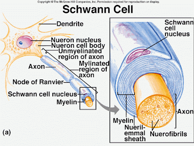
Schwann Cells/ Neurolemmocytes
Source

  • Schwann cells have the same function in the PNS that Oligodendrocytes have in the CNS, namely myelination.
  • The cell wraps itself around the axon of a neuron, insulating it, as seen below.  Pretty cool, eh?


Source on brainyinfo.com


Satellite Cells
  • Cushion neurons
  • Help control the environment of neurons and maintain synaptic integrity by insulating areas where there shouldn't be additional synapses on the neuron.


Tuesday, August 27, 2013

Plant Embryos

This post will be a running work in progress as I go through my Plant Biology class.  Here's what I've learned so far.


Source: stolaf.edu
The above picture (yes it's an actual photo of something under a microscope) is a good simple representation of the cotyledons of a dicot plant.  "Di" means "two", and "cot" refers to the cotyledons, so all embryonic dicots have two cotyledons.  Monocots then, naturally, have one cotyledon ("mono" means "one").
Simply put, cotyledons are embryonic leaves of the plant.

Here's a better detailed diagram:


Source on Britannica

Notice again that the dicot has 2 cotyledons.  Epicotyl will give rise to the shoot system and is above the cotyledons, while the hypocotyl is below the cotyledons and also contributes to the shoot.  This is easy to remember because "epi" means above or upon, while "hypo" means below.

The radicle gives rise to the root system.

Here's a fun picture showing a peanut.  The yummy part we eat is actually the cotyledons (leaves!).  I never knew that little nub in the middle that I sometimes discard was the actual plant in the making. :)


Key Terms in my own words (clicking on the term will take you to the wikipedia page for more info)  *PLEASE do not copy/ paste this to plagiarize homework assignments.  Read about it, rephrase in YOUR own words.  Don't let me be a source of academic dishonesty, even though I know a simplified list like this lends itself to that.  Please, please... be a smart cookie.
Cotyledon: embryonic leaf
Dicot: a category of plants which has 2 cotyledons in its embryonic stage
Monocot: a category of plants which has 1 cotyledon in its embryonic stage
Radicle: embryonic root.  First thing to emerge from seed.
Epicotyl: gives rise to shoot system, is above the cotyledons.
Hypocotyl: is below the cotyledons, contributes to the shoot system, adds height to the plant

Saturday, August 24, 2013

Long time no see. My excuse: boyfriend and Facebook

I'm very embarrassed to admit I have 6 posts in draft form.  Since I haven't posted since, what, March?... It's time I get my blogging juices flowing again by just putting SOMETHING live on the internet and then recommitting to posting more frequently (and to finishing those 6 posts!).

The interesting thing is I don't have readers that follow everything I post, it's more like a bunch of random people who come to learn about one particular thing I have blogged about and pops up on their search engine.  Which is great, I'm glad to be an educational source in any capacity.  But it does kind of lend itself to me being lazy.  People can still access my very popular posts anytime, so I don't feel like I'm letting anyone down or anything.  In fact, the posts I do like this one that doesn't have a specific scientific topic, usually don't even get read.

This blog has been my life, my love, my passion for almost 2 years, but the honest truth is - I got a boyfriend in March and since then my "free time" has no longer been used on blogging!  I have, however been doing lots of awesome real-life stuff.  Here, I'll put some pics to prove it! (Posted at the bottom.)

But I miss my blog and I feel I've been neglecting a huge part of "me", and I need to get back to it.

School starts Monday so this is a good chance to focus more on science and learning- my blog helps me do that by focusing my own studies and expanding on them.  Luckily I am taking NEUROSCIENCE this semester, and I'm so excited you don't even know!  So I bet you'll be seeing lots of posts related to that topic.

I think another reason for my recent blog laziness is that I got more involved on Facebook.  I found lots of really awesome science pages that post way cool stuff, so I've been getting my science fix that way.  Some of my favorites are: Science Is AwesomeGive a S*** About Nature, All Science All The Time, Nerds Do It BetterScience: The Magic of Reality, Science Alert, and lots more.  I get posts from probably 20 different places, it's awesome.  What I need to actually do is find out the info there, and then post it here!  I will try to start doing that and getting the info out to more people in Google search land.

So, here we go, another semester of awesome learning, coming right up!

Spelunking 3/14/2013

Holding a real human brain at The Leonardo 3/16/2013
Post on this


Hiking by Rock Canyon 3/29/2013

Hiking in the rain up Dry Canyon - found a deer carcass
Post on this

Visit to the Zoo - a friend of my boyfriend gave us a personal animal show :)

Zoo 4/29/2013

Backpacking up Dry Canyon 5/11/2013

I built a fort on Mother's Day 5/12/2013


Collared Lizard on a waterfall hike 5/20/2013

First time River Rafting!  Woohoo!  5/20/2013

Hiking a little peak in Roosevelt

Touching a snake from the Bean Museum


A sunset hike up dry canyon with the boys

Climbing trees at the duck pond
Found this weird animal up the tree

While camping, found this great part of the river to play in, in Spanish Fork 6/2/2013 


Dinosaur National Monument! 6/30/2013
I love bones!

Rain in Denver, Colorado 7/1/2013

Backpacking in the Rocky Mountains in Denver 7/3/2013


I got to be a camp counselor at Nature High Summer Camp! 7/8 - 7/13/2013
Post in the works


My boyfriend made a Portal cake for my birthday! :D 7/13/2013

We finished the summer off with a visit to the hospital (appendectomy).  At least a visit from the Kid History guys brightened things up.