Showing posts with label zool2420. Show all posts

Friday, April 4, 2014

Blood Clotting

Blood clotting or coagulation (also called hemostasis), is complex, but we want to try to understand this in "big picture" form first off.

When an injury occurs in a blood vessel, here are the steps we go through.


Classes.midlandstech.com

Platelets and Fibrin are the important things that we actually get as products of coagulation that seal up a wound.  But the activation of them is a complicated process.  Which is a good thing for your body!  If it were an easy reaction, we could get our blood spontaneously clotting on us and that would be BAD.

Here's another way to visualize this:

studyblue.com

In first forming the platelet plug, it's important to note that it's the exposed collagen fibers that are attracting platelets.  The normal, healthy state of things is to NOT have platelets sticking to blood vessels.  The ability to not stick is ensured by the blood vessel lining cells (endothelial) releasing prostacyclin to prevent platelet sticking.  But with damage, that isn't released and instead collagen causes platelets to stick.  This can be visualized here:

dc416.4shared.com

Now for the actual clotting part after the platelet plug, for now I have some videos.  If you want to cut right to the chase and a great explanation, watch the last video.


Videos
Here's a not fabulous animation but an animation nontheless...


This short clip I just found helpful to visualizing how the different factors work together to activate factor X then prothrombin, to activate thrombin which they refer to as the "thrombin burst" because it creates a large amount then creates a positive feedback loop to further increase the effect.


This is the best video I found!  I like how concise and understandable he makes it, looking at the big picture and working backward from there.  Hope it's helpful for you too.

All clotting factors are made by the liver, except 8 and PAF3. (Platelet activating factor is made by white blood cells.)

Wednesday, April 17, 2013

Kidney Physiology


Great overview


Urine Production Video - Osmotic Gradients

This is kind of fast, but it shows the counter-current diffusion of water and ions from the Nephron loop and vasa recta.

.


Here's a fun video - why coffee and alcohol make you pee more.


Wednesday, April 10, 2013

Respiration, Oxygen, and Hemoglobin

By decreasing pressure inside the thoracic cavity, we are able to inspire air.  By contracting the diaphragm, intercostal muscles, and others, we increase the size of the thoracic cavity, which in turn lowers the pressure to the point that it is lower than the outside air, causing a pressure gradient so the air rushes into the lungs.  To exhale, it is the opposite- relax the muscles, decrease the space in the cavity, which increases the pressure inside until higher than outside, so the air rushes out to where the pressure is lower.

This video is a good summary.  The embedding doesn't work, so you'll have to click the link instead.  Just left the embed on there for the picture. :)

Partial Pressure
Total Pressure of air can be broken into the partial pressures of all the gases contained in that air.  For instance, if the atmospheric pressure is at 760 mm Hg, and 20% of that is Oxygen gas, 80% Nitrogen gas, then the partial pressure of O2 would be 20% of 760 = 152 mm Hg.  Partial Pressure of N2would be 80% of 760 = 608 mm Hg.
This illustrates how increasing the pressure, as in the B picture, causes more gas to diffuse into the liquid.  A simple way to think about it is just that the higher the partial pressure of that gas, the less space it has to bounce around in the air, so more of it will end up in the liquid.

Here's a long video on partial pressure and gases getting into solution, I didn't watch the entire thing yet but it looks like a good detailed explanation for those who feel they could use more information.

Wednesday, April 3, 2013

Regulation of Blood Pressure

Blood Pressure is a very important aspect of homeostasis.  So naturally the body has many methods of monitoring and adjusting blood pressure to keep within a range that is functional for life.


Baroreceptor Reflex
Baroreceptors are located in the aortic arch and carotid sinuses (pictured below).  These are mechanoreceptors that respond to stretching of the arteries in response to blood pressure.


When these receptors are stimulated, nervous impulses are sent to the medulla oblongata of the brain where the reflex centers for vasomotor, cardiac, and respiratory are.


Wednesday, March 20, 2013

Heart

.

.

Here is a pretty good visual to show what gap junctions and desmosomes are.  The heart has both.

Tuesday, March 5, 2013

Muscle Contraction

Muscle Contraction is a very cool, amazing process.

Within the myofibrils, this is what's happening:
Source
Here's the explanation of the picture from the source's website:
a. Without calcium ions (in relaxed state). Because tropomyosin completely covers the sites (indicated in blue) of actin binding to myosin, myosin cannot bind to actin.
b. When calcium ions released from excited muscle cells bind to troponin, the binding sites of actin are exposed owing to the shift of tropomyosin; however, the extent of the shift is not sufficient for myosin to bind to actin.
c. When small amounts of myosin heads bind to the binding sites of actin under state b, tropomyosin shifts further, resulting in the complete exposure of the binding sites of actin.
d. Because the binding sites of actin are completely exposed, many myosin heads can bind to actin, which enables the generation of a large muscle contraction power. 


I love that it is showing the myosin heads (green globs) all the way around the actin.  Most of the time we look at it as a very 2-dimensional thing, but when you look at the picture below, you can see that it is a very 3-dimensional process, with myosin heads able to bind all the way around.  Each of the small dots represent actin, the bigger ones are myosin (seen on the ends of the myofibrils).
Source

Keep in mind, myofibrils are organelles within a muscle cell.  So the sarcoplasmic reticulum is the muscle version of ER, wrapped around each myofibril.  Stuff is diagrammed 2D to make it simpler and more understandable.  Once you understand it, it's cool to look back and imagine the bigger picture.


Videos!

Love to weed through the options and find a few good ones for you, this time I've included a song, rap, and skit!


Very short and sweet, this is just a good little animation to show how the filaments slide past each other, with the myosin heads moving along at different times.

One recommended by a student.  It says it's for invertebrates, but looks like it's accurate for us as well.


Hahaha, this is a fun one.  A song/rap explaining the whole process.  I probably like it because the background music is Daft Punk (they did the music for Tron Legacy).  Anyway!  Maybe it's helpful for someone, and it's kinda fun to listen to.


This skit is a great idea.  It's hard to read their signs some times, but this was a fun little video and it might help a couple things click for you like it did for me.  I want to get a bunch of people to act something like this out some day! :P  (Don't worry, no plans to make my study sessions do this...)


The singing on these videos often leaves much to be desired!  But the info in this one is pretty good...

Here's Dr. Ashworth's explanation she gave permission to share.  Thanks Jessie for recording and sending it.


Enjoy. :)

Wednesday, February 27, 2013

Nervous System

The nervous system is divided into Central Nervous System (brain and spinal cord) and Peripheral Nervous System (nerves, ganglia, etc).
Here's a little chart, courtesy of a great Physiology student:

Peripheral Nervous System Central Nervous System
nerves tracts
ganglia nuclei
Schwann cells oligodendrocytes


Part of the Peripheral Nervous System (PNS) is the involuntary part, consisting of efferent neurons controling visceral organs.  This is called the visceral nervous system, but more commonly known as:
Autonomic Nervous System
Autonomic = Automatic = Involuntary
Sympathetic & Parasympathetic divisions.  Here's a silly little video that shows the functions of each.


Sympathetic Division
Location: Thoracic and Lumbar regions of spinal cord

3 Kinds of pathways


Parasympathetic Division
Location: Brain, and Sacral region of spinal cord


Receptors


This is super fun, talks about the brain, gets deep and slightly philosophical, and mentions reflexes.  It's all about the brain! :)


Monday, February 18, 2013

Reflexes

Reflex Arcs involve the spinal cord and leave the brain out of the picture, in order for the compensatory action to happen very quickly and avoid tissue damage.  Here is a short and sweet explanation:


Thursday, February 14, 2013

Glucose Metabolism - Glycolysis through Kreb's Cycle

I was trying to think of a way to model these processes for the benefit of kinesthetic learners, so this is what I came up with using these cool toys my kids got from the kids' meal at Wendy's.  I hope this will help people visualize this and understand where all the Carbons from glucose go, and why on earth we care about all those NAD, NADH, FAD, and FADH2.


Here are the symbols for this model.  Purple= Carbon, Blue= Hydrogen, Red=NAD, Green=FAD, Orange=CoA enzyme


Below is a VERY simplified version of a glucose molecule.  This shows the 6 carbons because we want to see what happens to all these carbons.  There should be 12 Hydrogens and 6 Oxygens as well but for simplicity, I have only the carbons and 2 Hydrogens to illustrate one round of Redox reactions in glycolysis.

Glycolysis
Here we start with a glucose molecule and we will break it down.  For this demonstration I am ignoring the ATP, but just note that 2 ATP's go in to Glycolysis and you get 4 ATP out (so, a net production of 2 ATP).

Here are the reactants, or what goes in to glycolysis:


Next, we see that the glucose is broken apart (in half, basically)

And the NAD's come in to take the Hydrogens off to become NADH.  FYI, the glucose was "oxidized" because the electrons were taken off it, and the NAD was "reduced" because it got the electron (the hydrogen).  So, this is a Redox (Oxidation-Reduction) reaction.

Now we have two 3-Carbon structures, and two NADH's


The 3-Carbon moleucles are Pyruvate.

That's the end of glycolysis.  The NADH's are sent on to the electron transport chain at this point, so they leave.  Next the Pyruvates will be taken on and processed further in the next process, sometimes called the "Linking Step".  We will follow one pyruvate - keep in mind this process would happen twice for each glucose molecule since it splits in half into 2 pyruvates.

Oxidation of Pyruvate to Acetyl CoA (aka "The Linking Step")
So here is the Pyruvate going into the linking step.  By the way, this is the point at which metabolism has entered the Mitochondria.  Glycolysis takes place in the cytoplasm, and then pyruvate enters the mitochondria and that is where the rest of the processes take place.


I didn't do an intermediate picture on this one, sorry about that.  The NAD comes and harvests another Hydrogen (which is not shown on my simplified pyruvate), CoA is added and one carbon is removed and disposed of as Carbon dioxide.  Here are the products, notice the 3 Carbons accounted for:


And, we see here where each of the products are headed now:
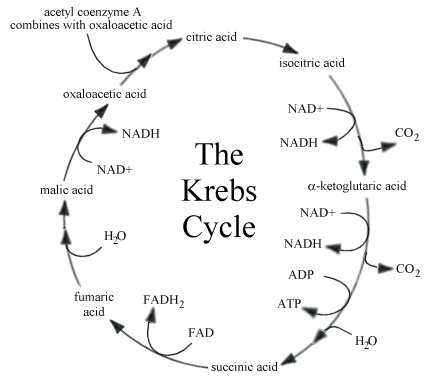
Kreb's Cycle
Then we follow the Acetyl CoA into the Kreb's Cycle, here are the reactants:


I didn't attempt to show all the steps of the Kreb's cycle here, but there are many redox reactions taking place, turning NAD and FAD into NADH and FADH2.  Here's a diagram of the cycle, if you really want to see it:


 Here you see the products, and see all the carbons are accounted for (2 Carbons in, 2 Carbons out):


And, here are the other products, which are the electron carriers that then deliver it over to the electron transport chain:

Also, note that 1 ATP is made during the Kreb's cycle which is not illustrated.

See this post on the Electron Transport Chain for some great videos about what happens there.  NADH and FADH2 are the carriers that drop off the electrons to run the electron transport chain, which harvests 30+ ATP per glucose molecule.  That is why we care so much about harvesting Hydrogens from glucose so we can make NADH and FADH2 and send them on their merry way to the electron transport chain to make ATP.  ATP is the energy that cells use to do pretty much everything!  If you stop making ATP, you die.  End of story.


So, to sum up - see if you can go through and account for all the Carbons, Hydrogens and Oxygens of glucose (glucose is C6 H12 O6).  You'll see that 6 carbons go in, and 6 carbons come out.  (Remember that each glucose molecule becomes TWO pyruvates, so you would double the numbers from that point on.)

Also, 6 oxygens go in, and how many come out?  Yup, 6.  Go back and check if you want to see for yourself. :)

Now, how about the Hydrogens?  Go through and count to see how many are harvested.  Really, go look and figure it out...


....
Did you come up with 14?  Did you count wrong?  NO!  It is 14.  But there are only 12 on glucose right?  What's the deal?  The reason is we actually have one molecule of water (H2O) that goes in the Kreb's cycle during one of the steps (it's added to fumaric acid to make malic acid), and those are then harvested off.  So there are the extra 2 hydrogens!  :)  It's a beautiful thing.

P.S. if you notice on the diagram of Kreb's, it shows 2 H20 going in, but one of them does come back out, as shown on the more detailed diagram below.  2 water molecules in, 1 back out, that's a net of 1 H20, so there are your extra 2 Hydrogens still.  I circled the H20 for you to see:
Source

Hope that helps you understand and visualize metabolism better!  I'd love if you would leave a comment, and follow this blog if you feel so inclined.  Thanks, and happy learning. :)

Thursday, February 7, 2013

Action Potentials Up Close


Here is a pretty nice video showing action potentials and how they are propogated without decreasing.


Another one which shows the voltage-gated Sodium and Potassium channels:


This animated image is a bit difficult to follow because it's so fast, but seems like it could be helpful to someone, especially if you know how to slow it down (which I don't- if you know, please teach me!).  So here it is.
Source


Here are a few various visualizations I liked

Source - awesome nervous system/eye/ear post!

Source


Source - great post with lots of details!

The following images were all from this excellent source: http://mikeclaffey.com/psyc2/notes-neurons.html

First, we see the steps of the action potential and what is going on with the various channels:
Source


Next, here is a nice depiction of how the action potential moves down the axon and why impulses travel one-way.

Source


Lastly, we see how saltatory conduction works.  This is the fastest way to conduct an impulse because it gets to "skip" all that length of the membrane on the axon that is myelenated.

Source


There you are, folks, another deeper view into the amazing world of Action Potentials and your nervous system...isn't this awesome!?

(If you missed the first post, here is a link to it)
Action Potentials - What Make Your Brain Work

Wednesday, February 6, 2013

Membrane Potential

Membrane Potential is a separation of charges across a membrane.  In other words, it is positive on one side and negative on the other.

You could think of the "potential" as potential to do work.  When you have concentration gradients of ions, that is potential energy, like the elevator analogy.

Wikipedia

I haven't taken the time to actually watch these, but there are several videos on Khan Academy going over this and may be a great resource:
https://www.khanacademy.org/science/healthcare-and-medicine/heart-depolarization/v/membrane-potentials---part-1


At 1:57-3:00 of this video, it discusses chemical/ concentration gradient and electrical gradient and how they get to equilibrium (resting membrane potential for potassium).


Thursday, January 31, 2013

Metabolic pathways

Overview
Source


.
Source
Energy from Carbohydrates
1. Glycolysis (breaks 1 glucose into 2 pyruvate)
Source

2. Pyruvate to Aceytl co-A
Source


3. Acetyl co-A into the Kreb's cycle
Source
4. FADH2 and NADH go into electron transport chain for oxidative phosphorylation (see post on this topic)

Energy from Proteins
1. Break into Amino Acids (hydrolysis)
2. Transamination: transfering the amino group to a carrier (amino acceptor)
Source
3. Deamination: take the amino group OFF and send it to be made into urea.  What's left over can be put into the Krebs cycle (alpha-ketoglutarate is one of the steps of Krebs).
Source
4. Can go straight from Amino Acids to Acetyl co-A then put that into Kreb's cycle
5. Can synthesize glucose out of amino acids when needed, which can then be used or stored as above in the section on carbs


Energy from Fats
1. Triglycerides can be broken down into glycerol and fatty acids
2. Glycerol can be used to make glucose (gluconeogenesis) in the liver
3. Fatty acids can be oxidized into Acetyl-coA (fatty acid oxidation) to go into the Krebs cycle, and harvest energy from there
 
Source


Wednesday, January 30, 2013

Membrane Transport and Osmotic Pressure

This is a good video giving an overview of all the different kinds of membrane transport.  It also includes an explanation of the sodium-potassium pump which is important in membrane potentials, and it would be great to watch this to help prepare for that section! :)


Overview of types of transport:

Source


Simple Diffusion
Dissolved stuff wants to get evenly spread out.
Source


Facilitated Diffusion
This shows all again, but notice the 2 in the middle:

Source

Osmosis
If the membrane isn't permeable to the solute (ions etc), water (the solvent) is what moves to try to get things evenly distributed.  This is called osmosis.
Source
Tonicity

Source

Due to the differences in tonicity, osmotic pressure can cause cells to change because the water moves trying to equalize the solute-solvent ratios.  Here is the classic example of red blood cells in plasma.  The hypertonic/ isotonic/ hypotonic is referring to the plasma or solution, in relation to the inside of the RBC.
Source


Primary Active Transport
Something is pumped against its concentration gradient, using ATP as the energy source
Source


Secondary Active Transport
Something is pumped against its concentration gradient, using something besides ATP as the energy source.  In these examples, one ion is pumped with its gradient, and that energy is used to pump something else against its gradient.  Here are a few examples:
Source


Source
.


.