Showing posts with label cells. Show all posts

Sunday, September 7, 2014

Plant Cell Wall Synthesis

Plants have some things animals don't, including a cell wall surrounding their cells.

 
Source of picture


Primary Cell Wall

Here's a diagram of the primary cell wall, along with the middle lamella that lies between adjacent plant cells with their respective primary cell walls.  Also the regular-old plasma membrane lies internally to all that.
Link to source
The middle lamella is made of pectins which are the perfect sticky thing to attach a primary wall made of cellulose microfibrils to!  The primary wall also has some other stuff to hold it together in a nice meshy business.

Synthesis of Primary Cell Well
This is the coolest part...

When a cell splits and becomes two cells, a new cell wall must be built between them.  I'll go into the details of the cytokinesis itself in another post, but after that is done, all that is there is is a middle lamella (again, made of pectins), and a plasma membrane on either side.  How does the primary wall end up BETWEEN the plasma mebrane and the middle lamella?!?

Cellulose synthase, that's how.  And it's brilliant.  In the plasma membrane, there is a complex of proteins embedded that make cellulose.  They look like little rosettes, like the ones depicted in blue, below:


The cellulose microfibrils get put together and come out of the external end of the rosettes (closer to the middle lamella).  The long cellulose molecules that strengthen the primary wall adhere to the middle lamella, add some cross-linking stuff (pectin, glycans) and there you have it.

The cool part is those rosettes actually move through the plasma membrane, (like wading through mud) guided by microtubules which are on the internal side of the plasma membrane, leaving the trail of cellulose as it goes.

Here are some other diagrams of how this works.

In this one, the blue arrows indicate the direction the rosettes are "wading" through the plasma membrane, "walking" along the orange microtubules beneath.

This shows how the glucose subunits come in from the cytoplasm (purple circles) and are put together into the complex polymer of cellulose.  Again, the arrow shows the rosette is moving to the left, leaving a trail of cellulose to the right.
Source of image


And here we see the yellow plasma membrane cut away partly so we can see the rosettes that pass through and synthesize the microfibrils of cellulose.  Each section of the rosette is an enzyme in its own right that puts together the long chains from glucose, which is then wound together into larger and larger units.
Source of image


Here's what the structure of cellulose looks like broken down, so you can see it's a complex, tightly packed polymer.
Source of image

The strands of cellulose are arranged pretty randomly in a primary cell wall.  The cellulose synthases don't move very quickly, so the cellulose that is spit out goes around rather randomly, much like squeezing a bunch of toothpaste out of a tube- it goes every which way.


Secondary Cell Wall Synthesis
The secondary cell wall is lain down internally to the primary cell wall.  It is thick and has 3 layers that are put down one at a time.  Each layer has all its cellulose going parallel to each other.  But the layers each have different directions/ orientations than one another, as seen in the bottom part of this diagram:

Source of image

This provides a lot of extra strength, because it is protecting against compression, stretching, tension, etc. in all directions once you have all 3 layers put down.  When the cellulose rosettes are laying down cellulose for a secondary cell wall layer, they move more quickly and in regular, straight lines.

That's all she (I) wrote.  Stay curious!

Thursday, January 2, 2014

Theodore Schwann

Theodore Schwann (many sources also spelled his name Theodor) was a man who lived in the 1800's and is remembered as a great scientist.  His actual scientific career only lasted five years!  And yet, in that short time (1834-1839), he made many very important discoveries in Biology and Physiology.


Source
Cell Theory
Shortly after German Botanist Matthias Schleiden discovered that plants are made of cells, Schwann discovered that animals are as well (Source).

Alcoholic fermentation
He hypothesized that fermentation wasn't a reaction with nitrogen in the air as people then believed, but that tiny organisms (yeast) were eating the sugars of the fruit and then excreting carbon dioxide and ethanol.  People in his day noticed yeast multiplied with fermentation, but he was the first to suggest they were the thing doing the fermenting, rather than just a byproduct of it.  He also stated that yeast were living plant-like organisms, firmly establishing fermentation as a biological process.  He was publicly ridiculed for this hypothesis which came at the end of his short career and probably helped lead to its demise, as he could no longer get funds or promotions to continue doing scientific research.  (Paraphrased from The Other Brain, by R. Douglas Fields, 2009, and Wikipedia- Theodore Schwann.) 

Digestion
He suspected that hydrochloric acid wasn't the whole story with digestion.  He discovered Pepsin, the enzyme that helps break down proteins (The Other Brain). He also showed that bile is essential to digestion, and coined the term "metabolism" describing chemical changes in animal tissue (Source).

Schwann cells
The myelinating cells in the Peripheral Nervous System are his namesake, since he first described them.  He studied nerves, noticing they had little dew-drop like structures running the length of them.  He thought that a neuron being built during development must join with other cells to create the long axon by fusion, and the Schwann cells were remnants of this process from those fetal cells.  Although his speculation was incorrect, these cells still carry his name.

Source
There's a brief summary of a great scientist's work.  I read about him in the book I've currently got my nose in (and hoping to finish before school starts back up), The Other Brain, by R. Douglas Fields.  It's all about Neuroglia, aka glia, glial cells.  To read an overview of all the types of Glia, go to this post: http://biogeonerd.blogspot.com/2013/08/neuroglia.html

Tuesday, September 3, 2013

Plant cells

The pictures on this blog post are all my own, taken through a microscope, sorry not all of them are the best quality.  But at least I don't have to collect links to sources!

Mitochondria- the largest dark dots are the nucleus, but you can see lots of little dark purple spots which are the mitochondria.  This is from a prepared slide of an onion root with a special stain for seeing mitochondria, and a brown stain for cell walls.

Chloroplasts
These look quite blurry, it's just because there are multiple layers of cells superimposed on one another.  This was a hand mounted water weed leaf.  All the little green speckles everywhere are chloroplasts- the organelles which house thylakoids for conducting photosynthesis. :)


Chromoplasts
These are like chloroplasts, but they are not green.  They are still colored though, hence the name chromo ("color") plast.  You can make out the little orange specks from a carrot:

And tiny red specks from a tomato:

Amyloplasts- these are a type of plastid that contains starch storage.  The tiny purple granules are the ones.  They're only purple because a stain was added to this potato so you could actually see it.


Thursday, February 14, 2013

Glucose Metabolism - Glycolysis through Kreb's Cycle

I was trying to think of a way to model these processes for the benefit of kinesthetic learners, so this is what I came up with using these cool toys my kids got from the kids' meal at Wendy's.  I hope this will help people visualize this and understand where all the Carbons from glucose go, and why on earth we care about all those NAD, NADH, FAD, and FADH2.


Here are the symbols for this model.  Purple= Carbon, Blue= Hydrogen, Red=NAD, Green=FAD, Orange=CoA enzyme


Below is a VERY simplified version of a glucose molecule.  This shows the 6 carbons because we want to see what happens to all these carbons.  There should be 12 Hydrogens and 6 Oxygens as well but for simplicity, I have only the carbons and 2 Hydrogens to illustrate one round of Redox reactions in glycolysis.

Glycolysis
Here we start with a glucose molecule and we will break it down.  For this demonstration I am ignoring the ATP, but just note that 2 ATP's go in to Glycolysis and you get 4 ATP out (so, a net production of 2 ATP).

Here are the reactants, or what goes in to glycolysis:


Next, we see that the glucose is broken apart (in half, basically)

And the NAD's come in to take the Hydrogens off to become NADH.  FYI, the glucose was "oxidized" because the electrons were taken off it, and the NAD was "reduced" because it got the electron (the hydrogen).  So, this is a Redox (Oxidation-Reduction) reaction.

Now we have two 3-Carbon structures, and two NADH's


The 3-Carbon moleucles are Pyruvate.

That's the end of glycolysis.  The NADH's are sent on to the electron transport chain at this point, so they leave.  Next the Pyruvates will be taken on and processed further in the next process, sometimes called the "Linking Step".  We will follow one pyruvate - keep in mind this process would happen twice for each glucose molecule since it splits in half into 2 pyruvates.

Oxidation of Pyruvate to Acetyl CoA (aka "The Linking Step")
So here is the Pyruvate going into the linking step.  By the way, this is the point at which metabolism has entered the Mitochondria.  Glycolysis takes place in the cytoplasm, and then pyruvate enters the mitochondria and that is where the rest of the processes take place.


I didn't do an intermediate picture on this one, sorry about that.  The NAD comes and harvests another Hydrogen (which is not shown on my simplified pyruvate), CoA is added and one carbon is removed and disposed of as Carbon dioxide.  Here are the products, notice the 3 Carbons accounted for:


And, we see here where each of the products are headed now:

Kreb's Cycle
Then we follow the Acetyl CoA into the Kreb's Cycle, here are the reactants:


I didn't attempt to show all the steps of the Kreb's cycle here, but there are many redox reactions taking place, turning NAD and FAD into NADH and FADH2.  Here's a diagram of the cycle, if you really want to see it:


 Here you see the products, and see all the carbons are accounted for (2 Carbons in, 2 Carbons out):


And, here are the other products, which are the electron carriers that then deliver it over to the electron transport chain:

Also, note that 1 ATP is made during the Kreb's cycle which is not illustrated.

See this post on the Electron Transport Chain for some great videos about what happens there.  NADH and FADH2 are the carriers that drop off the electrons to run the electron transport chain, which harvests 30+ ATP per glucose molecule.  That is why we care so much about harvesting Hydrogens from glucose so we can make NADH and FADH2 and send them on their merry way to the electron transport chain to make ATP.  ATP is the energy that cells use to do pretty much everything!  If you stop making ATP, you die.  End of story.


So, to sum up - see if you can go through and account for all the Carbons, Hydrogens and Oxygens of glucose (glucose is C6 H12 O6).  You'll see that 6 carbons go in, and 6 carbons come out.  (Remember that each glucose molecule becomes TWO pyruvates, so you would double the numbers from that point on.)

Also, 6 oxygens go in, and how many come out?  Yup, 6.  Go back and check if you want to see for yourself. :)

Now, how about the Hydrogens?  Go through and count to see how many are harvested.  Really, go look and figure it out...


....
Did you come up with 14?  Did you count wrong?  NO!  It is 14.  But there are only 12 on glucose right?  What's the deal?  The reason is we actually have one molecule of water (H2O) that goes in the Kreb's cycle during one of the steps (it's added to fumaric acid to make malic acid), and those are then harvested off.  So there are the extra 2 hydrogens!  :)  It's a beautiful thing.

P.S. if you notice on the diagram of Kreb's, it shows 2 H20 going in, but one of them does come back out, as shown on the more detailed diagram below.  2 water molecules in, 1 back out, that's a net of 1 H20, so there are your extra 2 Hydrogens still.  I circled the H20 for you to see:
Source

Hope that helps you understand and visualize metabolism better!  I'd love if you would leave a comment, and follow this blog if you feel so inclined.  Thanks, and happy learning. :)

Wednesday, February 6, 2013

Membrane Potential

Membrane Potential is a separation of charges across a membrane.  In other words, it is positive on one side and negative on the other.

You could think of the "potential" as potential to do work.  When you have concentration gradients of ions, that is potential energy, like the elevator analogy.

Wikipedia

I haven't taken the time to actually watch these, but there are several videos on Khan Academy going over this and may be a great resource:
https://www.khanacademy.org/science/healthcare-and-medicine/heart-depolarization/v/membrane-potentials---part-1


At 1:57-3:00 of this video, it discusses chemical/ concentration gradient and electrical gradient and how they get to equilibrium (resting membrane potential for potassium).


Wednesday, January 30, 2013

Membrane Transport and Osmotic Pressure

This is a good video giving an overview of all the different kinds of membrane transport.  It also includes an explanation of the sodium-potassium pump which is important in membrane potentials, and it would be great to watch this to help prepare for that section! :)


Overview of types of transport:

Source


Simple Diffusion
Dissolved stuff wants to get evenly spread out.
Source


Facilitated Diffusion
This shows all again, but notice the 2 in the middle:

Source

Osmosis
If the membrane isn't permeable to the solute (ions etc), water (the solvent) is what moves to try to get things evenly distributed.  This is called osmosis.
Source
Tonicity

Source

Due to the differences in tonicity, osmotic pressure can cause cells to change because the water moves trying to equalize the solute-solvent ratios.  Here is the classic example of red blood cells in plasma.  The hypertonic/ isotonic/ hypotonic is referring to the plasma or solution, in relation to the inside of the RBC.
Source


Primary Active Transport
Something is pumped against its concentration gradient, using ATP as the energy source
Source


Secondary Active Transport
Something is pumped against its concentration gradient, using something besides ATP as the energy source.  In these examples, one ion is pumped with its gradient, and that energy is used to pump something else against its gradient.  Here are a few examples:
Source


Source
.


.

Electron Transport Chain & Oxidative Phosphorylation

This process is really confusing when not understood, and really phenomenal and awesome when understood.  Learn and geek out with me, if you will. :)
First, the electron transport chain (done by Virtual Cell):


Next, the explanation of ATP Synthase:


And a beautiful animation of these processes (see if you can point out the players):


I am so grateful that Virtual Cell and BioVisions have made these awesome videos!

Wednesday, January 23, 2013

Protein Synthesis

As a quick overview, you can see the processes of transcription, translation, protein synthesis and exocytosis in this video.  It's detailed and labelled, the pertinent parts for this post are at minute 4:30 to 5:30, roughly:

DNA --> mRNA --> tRNA --> Amino Acids

Transcription
Source: wikipedia
Source
Translation

Source
Here's a great video of this process:

Cell Organelles

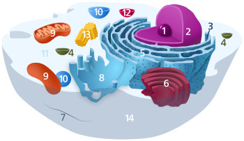
Here are some various drawings of an idealized animal cell, showing all the different types of organelles.  Different artists depict them slightly differently, so looking at many versions should be helpful.  If you click the "source" link under each picture, you will find many great websites with further information.  In some cases this will be necessary to be able to see the image.  My black background is messing some of these up, sorry about that.
Look at these to orient yourself, we will then go into detail on each organelle, and then have some unlabeled cell drawings so you can quiz yourself.  Enjoy!


Source


Source


Source


Source


Source


Organelles
Here are some closeups! :D

Cell membrane


Here is a good depiction of the various things embedded into a cell's plasma membrane (also referred to as the phospholipid bilayer):

Source


Nucleus

Notice the pores, which is where the mRNA (post-transcription) would emerge to go to a ribosome for translation.
Source

Mitochondria

Source


mitochondria
Source
Here is a labelled mitochondria.  Please note, Matrix and Cristae will come into play when you learn about metabolism.
Source
And, here is a really sweet video of a mitochondrion, showing some metabolism processes.  FYI Mitochondrion is singular, Mitochondria is plural.  (Link to the animator's website, for better quality, and links to other really cool videos: http://www.xvivo.net/powering-the-cell-mitochondria/)


Golgi

golgi body
Source


Cytoskeleton

Source

Identification Practice

Source
Source


Source: wikipedia

Key for wikipedia image (includes links to wiki pages of each):

Organelles:
1 Nucleolus
2 Nucleus
3 Ribosomes (little dots)
4 Vesicle
5 Rough endoplasmic reticulum
6 Golgi apparatus
7 Cytoskeleton
8 Smooth endoplasmic reticulum
9 Mitochondria
10 Vacuole
11 Cytosol
12 Lysosome
13 Centrioles within Centrosome
14 Cell membrane

The end