Showing posts with label plant cells. Show all posts

Thursday, October 2, 2014

Leaf Anatomy

Meristems
View of a shoot apical meristem with some leaf primordia.  Additionally, I have labelled the 3 primary meristems you can differentiate here.


Here's a nicotine leaf to show the two kinds of meristems specific to the leaf.  The leaf apical meristem becomes the midrib of the leaf, while the leaf marginal meristem is the blade of the leaf.


Epidermis
From a Sedum (stone crop)- you can see the "regular" epidermal cells (squiggly shaped) as well as the guard cells in various places surrounding stomata.

 Take a closer look:

Also here is a sunken stomate as seen in a Pinus leaf.  It is "sunken" because the guard cells are below the level of the epidermis which helps protect against dessication (drying out).


Mesophyll
Mesophyll just means middle of the leaf.  It is the term for the ground tissue in the leaf.  There are two types by shape: palisade and spongy.
You can see both of these well in a pine leaf:

We also term a leaf to be unifacial or bifacial based on the arrangement of the mesophyll.  Bifacial is if there is spongy on one side, and palisade on the other.  Unifacial is either the same type throughout, or it makes a kind of sandwich with the same kinds on either side and something different in the middle.  You can see this type of unifacial in a Dianthus (carnation) leaf:
(The red rod-shaped parts are palisade, the lighter stained area in the middle with a lot of spaces is spongy.)

Pine anatomy
Some things we learned specifically with pine needles (I'm not sure if they also apply to some other plants, sorry) are the resin duct with epithelium, hypodermal sclerenchyma, and transfusion tissue.
Some of these are labeled:


The outermost layer surrounding the resin duct is the epithelium.  Hypodermal sclerenchyma is below ("hypo") the outermost layer of cells or dermis.  These have thick secondary cell walls which add to the strength of a pine needle, as you have probably tested yourself many times when you got poked with one.  In this repeat picture you can see the hypodermal sclerenchyma with the thick red-stained walls, on either side of the sunken stomate.

Bundle Sheaths
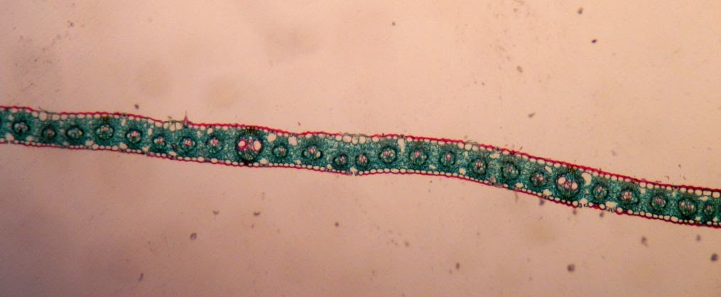
Bundle sheaths are different in C3 plants and C4 plants.
Here, in a C3 grass (Poa), you see their regular bundle sheaths:


Closer.  Just looks like a blank set of cells surrounding the vascular bundle.

But in a C4 plant, like this Zea corn, the sheaths have what is referred to as "Kranz anatomy".  Kranz is German for wreath, and you can see they are rather leaf-like in the following examples. There are two orientation pictures, then it zooms in one a single bundle sheath so you can see the Kranz anatomy.


Leaf Abscission Zone
When a plant loses its leaves, it prepares for this by creating an abscission zone so the leaf can easily fall off without damaging any of the other tissue.  Layers of cells secrete suberin (or is it subirin?) for protection (and are called suberized cells), while the next outermost layer of cells is pre-programmed to break easily, as it were.  The weak layer is called the separation layer.


Whew, that's a lot of leaf anatomy!  Stay curious.


P.S. There are no picture source references cause I took all these with my camera (through the microscope in Botany lab) and did the labeling myself.

Sunday, September 7, 2014

Plant Cell Wall Synthesis

Plants have some things animals don't, including a cell wall surrounding their cells.

 
Source of picture


Primary Cell Wall

Here's a diagram of the primary cell wall, along with the middle lamella that lies between adjacent plant cells with their respective primary cell walls.  Also the regular-old plasma membrane lies internally to all that.
Link to source
The middle lamella is made of pectins which are the perfect sticky thing to attach a primary wall made of cellulose microfibrils to!  The primary wall also has some other stuff to hold it together in a nice meshy business.

Synthesis of Primary Cell Well
This is the coolest part...

When a cell splits and becomes two cells, a new cell wall must be built between them.  I'll go into the details of the cytokinesis itself in another post, but after that is done, all that is there is is a middle lamella (again, made of pectins), and a plasma membrane on either side.  How does the primary wall end up BETWEEN the plasma mebrane and the middle lamella?!?

Cellulose synthase, that's how.  And it's brilliant.  In the plasma membrane, there is a complex of proteins embedded that make cellulose.  They look like little rosettes, like the ones depicted in blue, below:


The cellulose microfibrils get put together and come out of the external end of the rosettes (closer to the middle lamella).  The long cellulose molecules that strengthen the primary wall adhere to the middle lamella, add some cross-linking stuff (pectin, glycans) and there you have it.

The cool part is those rosettes actually move through the plasma membrane, (like wading through mud) guided by microtubules which are on the internal side of the plasma membrane, leaving the trail of cellulose as it goes.

Here are some other diagrams of how this works.

In this one, the blue arrows indicate the direction the rosettes are "wading" through the plasma membrane, "walking" along the orange microtubules beneath.

This shows how the glucose subunits come in from the cytoplasm (purple circles) and are put together into the complex polymer of cellulose.  Again, the arrow shows the rosette is moving to the left, leaving a trail of cellulose to the right.
Source of image


And here we see the yellow plasma membrane cut away partly so we can see the rosettes that pass through and synthesize the microfibrils of cellulose.  Each section of the rosette is an enzyme in its own right that puts together the long chains from glucose, which is then wound together into larger and larger units.
Source of image


Here's what the structure of cellulose looks like broken down, so you can see it's a complex, tightly packed polymer.
Source of image

The strands of cellulose are arranged pretty randomly in a primary cell wall.  The cellulose synthases don't move very quickly, so the cellulose that is spit out goes around rather randomly, much like squeezing a bunch of toothpaste out of a tube- it goes every which way.


Secondary Cell Wall Synthesis
The secondary cell wall is lain down internally to the primary cell wall.  It is thick and has 3 layers that are put down one at a time.  Each layer has all its cellulose going parallel to each other.  But the layers each have different directions/ orientations than one another, as seen in the bottom part of this diagram:

Source of image

This provides a lot of extra strength, because it is protecting against compression, stretching, tension, etc. in all directions once you have all 3 layers put down.  When the cellulose rosettes are laying down cellulose for a secondary cell wall layer, they move more quickly and in regular, straight lines.

That's all she (I) wrote.  Stay curious!