I have blogged quite a bit about Action Potentials which are the way in which a nervous impulse travels down an individual cell. (For your reference: Action Potentials and Action Potentials Up Close).
Here I will summarize what it takes for a message to be passed from one neuron to another. If you are interested in information about the specific channels that make neuron communication possible, see this post on Neurons.
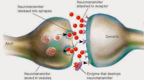
When an action potential - the electrical signals that neurons use to send messages - reaches the end of an axon, the electrical message must become a chemical message in order to cross the synapse. Neurons are connected to each other by a synapse which consists of the axon of the presynaptic neuron (the one sending the message), and the dendrite or cell body of the postsynaptic neuron (the one receiving the message), and the space between them called the synaptic cleft. Chemicals have to be sent across this space for the message to continue on. The chemicals used are neurotransmitters.
Here's a simple diagram of a synapse:
For a little more information and overview of neuron communication, here is a more detailed diagram:
When the electrical signal reaches the end of the axon, a special thing happens with Calcium entering the cell which allows those vesicles full of Neurotransmitter chemicals to exocytose. That's a fancy word meaning the vesicle smooshes into the cell membrane, kicking its contents outside the cell- into the synapse. Those neurotransmitters are then free to act on the postsynaptic cell dendrites or body to continue the message along!
But how did those vesicles of neurotransmitter actually GET there? The cell factories that make neurotransmitters which are mostly made of protein, are waaaaaay back at the cell body, but it has to be released at the terminal button of the axon! Axons can be very long, like so:
Neurotransmitter vesicles are continually made by the neuron (each neuron only makes one particular type of neurotransmitter), and sent down the axon. They get there the same way you would travel a long distance- on a "highway" of sorts.
Here is a great animation of the whole neuron communication process, and you will see vesicles coming down to the end of the axon.
So, the cool thing is those vesicles are literally "walked" down the microtubule highway. In my favorite video, you can see this happen:
Lastly, I found this funny little video about the life of a motor protein (the alien-looking guy that walked the vesicle down the microtubule). Enjoy!
P.S. While searching for images, I ran across this amazing blog, so here is a great reference on the brain and neuroscience, explained in simple terms in much the same way I try to do my own blog. The Brain Geek
Stay curious!
-Julie
Tuesday, August 26, 2014
Neuron Communication
Labels:
biology,
brain,
neurons,
neuroscience,
physiological psychology,
physiology,
synapse
Plant Biology Lab 1
Today we simply went over microscope procedures and practice making wet mounts of various plant parts.
Here are several pictures of a flower petal. The big circles of pink are just cells whose giant vacuoles are full of pigment. The 3 pictures are all of the same view and I just found it interesting that they look SO different, just with slight adjustments of the microscope or even the angle or temperament of my camera shooting through the eye piece.
This is a side view of a leaf. On the left (under side I believe) is were stomata would be. The circular thing in the middle of the picture that I *thought* was a stomate is actually a vascular bundle!
Again, different settings can change the coloring completely.
Here is water leaf, and we got some bonus little brown flecks, which our professor said are likely diatoms that were in the water with the plant.
The large purple splotches are large diatoms! The details inside the cells here is beautiful.
Dr. Robbins also showed us this neat video of chloroplasts doing "cyclosis". Here's the explanation of what that is on the description of this video on YouTube:
Rapid cycling of cytoplasm - cyclosis is seen here as circling of chloroplasts in cells of Elodea canadensis. Chloroplasts normally move in the cell to adapt to changing light conditions; here they are exposed to much too strong light (under the microscope) and they want to move away from it, but in the end they just circle round and round.
So there we go, a simple introduction to our botany lab. More exciting stuff will come in the weeks to follow. :)
Subscribe to:
Comments (Atom)


.JPG)


